Session Information
Date: Tuesday, November 14, 2023
Title: (2387–2424) Vasculitis – Non-ANCA-Associated & Related Disorders Poster III
Session Type: Poster Session C
Session Time: 9:00AM-11:00AM
Background/Purpose: Polymyalgia rheumatica (PMR) is associated with giant cell arteritis (GCA) in 16 to 21% of cases. This raises the question of a pathophysiological continuum between PMR and GCA, especially since a study reported mature arterial wall dendritic cells (DC) in patients with GCA or PMR. There are 3 main types of DC: plasmacytoid DC (expressing CD123), conventional DC (cDC) expressing CD141 (cDC1) or CD1c (cDC2), and monocyte-derived DC (mo-DC) expressing CD209 and CD14.This study aimed to identify, localize and characterize the phenotype of arterial DC in PMR and GCA.
Methods: Using temporal artery biopsies (TAB) from patients with PMR, GCA and healthy controls, bulk RNA-sequencing and RT-PCR analyses were performed to assess the level of expression of myeloid DC genes (CD141, CD1C, CD209, ITGAX), DC maturation genes (CD83, CD80/86, CCR7) and mature DC chemokine expression (CCL18, CCL19, CCL20, CCL21). Expression of markers of DC lineage (CD209), DC maturation state (CD83 and CCR7) and DC origin (CD14, CD68, CD1c, CD141) were also studied in TABs by immunofluorescence (IF).
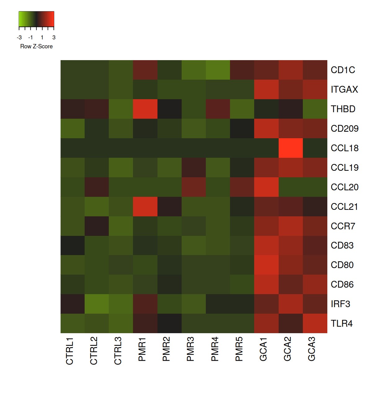
Results: Forty-three patients were included (14 GCA, 15 PMR, 14 controls). TAB from GCA patients were characterized by a strong mature DC signature with high level of expression of DC associated genes (CD1C, ITGAX [coding CD11c] and CD209 [coding DC-SIGN]), DC maturation genes (CD83, CCR7, CD80/CD86) and DC chemokine genes (CCL18, CCL19, CCL20). In PMR arteries, the DC signature was more heterogeneous: a few arteries expressed CD1C and CD141 but none expressed DC maturation genes. Healthy arteries were characterized by the absence of expression of DC associated genes and mature DC genes (figure 1). In GCA arteries, IF analysis revealed that the three arterial layers were heavily infiltrated by CD209+ cells. These cells also expressed CD14 and often CD68, thus fitting with mo-DC (CD209+CD14+CD68–) or macrophages (CD209+CD14+CD68+). Some of these cells expressed maturation markers of DC (CD83 and CCR7). However, no CD1c or CD141 cells were found in the arterial wall by IF. In PMR and control arteries, no DC were found by IF. Transcriptomic analysis revealed that GCA arteries expressed many genes involved in the differentiation of monocytes into mo-DC, including CSF2, CYBA, IRF4, AHR, WNT5A, and FZD2.
Conclusion: This work demonstrates the presence of mature CD209+CD83+CCR7+ DCs within the arterial wall in GCA but not in PMR or healthy arteries. The phenotype of these DCs mainly fits with mo-DCs. In PMR, the DC signature is more heterogeneous without expression of DC maturation markers.
Myeloid dendritic cell genes are defined as genes that characterize human dendritic cells: CD1C (coding CD1c), THBD (coding CD141), CD209, ITGAX (coding CD11c).
DC maturation associated genes are defined by the process in which antigen-activated dendritic cells acquire the specialized features of a mature conventional dendritic cell. Mature conventional dendritic cells upregulate the surface expression of MHC molecules, chemokine receptors and adhesion molecules, and increase the number of dendrites (cytoplasmic protrusions) in preparation for migration to lymphoid organs where they present antigen to T cells: CD83, CD80, CD86, CCR7, CCL18, CCL19, CCL20, CCL21, TLR4, IRF3.
To cite this abstract in AMA style:
ramon a, Greigert h, Richard c, cladière c, genet c, ciudad m, tarris g, martin l, arnould l, creuzot-garcher c, ornetti p, Audia S, boidot r, Maillefert j, Bonnotte B, Samson M. Characterization of Arterial Dendritic Cells in Polymyalgia Rheumatica and Giant Cell Arteritis [abstract]. Arthritis Rheumatol. 2023; 75 (suppl 9). https://acrabstracts.org/abstract/characterization-of-arterial-dendritic-cells-in-polymyalgia-rheumatica-and-giant-cell-arteritis/. Accessed .« Back to ACR Convergence 2023
ACR Meeting Abstracts - https://acrabstracts.org/abstract/characterization-of-arterial-dendritic-cells-in-polymyalgia-rheumatica-and-giant-cell-arteritis/

