ACR Meeting Abstracts

ACR Meeting Abstracts

  • Meetings
    • ACR Convergence 2024
    • ACR Convergence 2023
    • 2023 ACR/ARP PRSYM
    • ACR Convergence 2022
    • ACR Convergence 2021
    • ACR Convergence 2020
    • 2020 ACR/ARP PRSYM
    • 2019 ACR/ARP Annual Meeting
    • 2018-2009 Meetings
    • Download Abstracts
  • Keyword Index
  • Advanced Search
  • Your Favorites
    • Favorites
    • Login
    • View and print all favorites
    • Clear all your favorites
  • ACR Meetings

Abstract Number: 1034

Changing Osteoarthritis Treatment Assessment Paradigms: Subchondral Bone Is a More Responsive Measure of Progression Than the Current Radiographic Standard

Michael A. Bowes1, Christopher B. Wolstenholme1, Devan Hopkinson1, Graham R. Vincent1 and Philip G. Conaghan2, 1Imorphics Ltd, Manchester, United Kingdom, 2University of Leeds, Leeds, United Kingdom

Meeting: 2012 ACR/ARHP Annual Meeting

Keywords: imaging techniques, Magnetic resonance imaging (MRI), osteoarthritis and radiography

  • Tweet
  • Click to email a link to a friend (Opens in new window) Email
  • Click to print (Opens in new window) Print
Session Information

Title: Imaging of Rheumatic Diseases: Magnetic Resonance Imaging, Computed Tomography and X-ray

Session Type: Abstract Submissions (ACR)

Background/Purpose: Radiographic joint space width (JSW) assessment, a surrogate for cartilage assessment, is the standard for structure modification trials of osteoarthritis (OA). However the subchondral bone is integral to OA progression and modern image analysis techniques allow accurate, automated identification of bone in MR images. The objective of this study was a comparison of the sensitivity of 3D bone area measures in MR images with minimum medial JSW in radiographs in all subjects in the Osteoarthritis Initiative dataset with definite medial OA over a 2 year period, representative of a typical OA clinical trial

Methods: 828 subjects with medial OA and MR images at baseline, 12 and 24 months, and available radiograph scoring were selected from the Osteoarthritis Initiative dataset; medial OA was defined as KL≥2 and presence of medial osteophytes. Femur, tibia and patella bones were automatically segmented from MRIs using active appearance models1. Anatomical areas were automatically identified within the model2 and were measured at each time-point. All regions of the articulating surface of the femur, tibia and patella were included in the analysis. Minimum medial joint space width (minMJSW) was provided by the OAI, using a semi-automated software method3. Sensitivity of both methods to change was assessed using the standardised response mean (mean/SD of change)

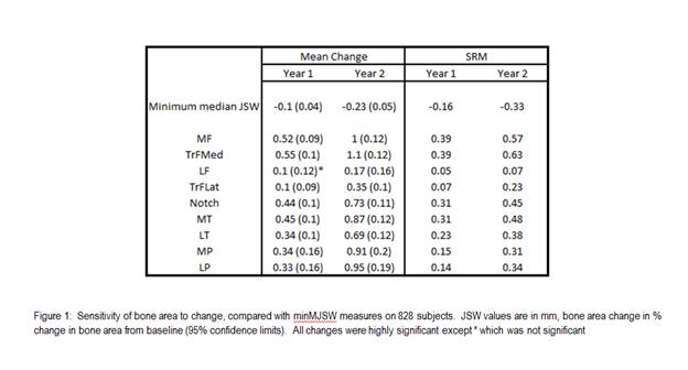
Results: Mean age (SD) of the case group was 62 years (8.8); BMI 29.7(4.8); KL 2.55(0.65); 35% females.  MinMJSW showed significant change at 12 and 24 months with SRM values of -0.16 and -0.33. Change in bone area was significant in all regions for all time-points, except the lateral femur at 12 months (see Figure 1).  Medial femur compartments provided the greatest sensitivity to change, with SRM values typically twice those of minMJSW.  Tibial compartments and the notch of the femur also showed higher SRM figures than minMJSW, while patellar compartments had comparable SRMs to the radiographic measure.  Only the lateral femoral compartments had lower SRMs than the minMJSW method.

Conclusion: Change in bone area was more sensitive than the minMJSW method in a number of knee compartments, particularly in the medial femur and tibia.  Measurement of bone change provided a more responsive tool for monitoring OA progression in a cohort selected for typical OA trial characteristics.

References:

1              Cootes, T. F, G. J. Edwards, and C. J. TaylorIEEE Trans Patt Anal Mach Intell 23.6 (2001): 681-85.

2              Williams, T. G., et al. Br.J.Radiol. 83.995 (2010): 940-48.

3              Duryea J, Li J, Peterfy CG, Gordon C, Genant HK.   Med Phys. 2000 Mar: 27(2): 580-91


Disclosure:

M. A. Bowes,

Imorphics Ltd,

1,

Imorphics Ltd,

3;

C. B. Wolstenholme,

Imorphics Ltd,

3,

Imorphics Ltd,

1;

D. Hopkinson,
None;

G. R. Vincent,

Imorphics Ltd,

3,

Imorphics Ltd,

1;

P. G. Conaghan,
None.

  • Tweet
  • Click to email a link to a friend (Opens in new window) Email
  • Click to print (Opens in new window) Print

« Back to 2012 ACR/ARHP Annual Meeting

ACR Meeting Abstracts - https://acrabstracts.org/abstract/changing-osteoarthritis-treatment-assessment-paradigms-subchondral-bone-is-a-more-responsive-measure-of-progression-than-the-current-radiographic-standard/

Advanced Search

Your Favorites

You can save and print a list of your favorite abstracts during your browser session by clicking the “Favorite” button at the bottom of any abstract. View your favorites »

All abstracts accepted to ACR Convergence are under media embargo once the ACR has notified presenters of their abstract’s acceptance. They may be presented at other meetings or published as manuscripts after this time but should not be discussed in non-scholarly venues or outlets. The following embargo policies are strictly enforced by the ACR.

Accepted abstracts are made available to the public online in advance of the meeting and are published in a special online supplement of our scientific journal, Arthritis & Rheumatology. Information contained in those abstracts may not be released until the abstracts appear online. In an exception to the media embargo, academic institutions, private organizations, and companies with products whose value may be influenced by information contained in an abstract may issue a press release to coincide with the availability of an ACR abstract on the ACR website. However, the ACR continues to require that information that goes beyond that contained in the abstract (e.g., discussion of the abstract done as part of editorial news coverage) is under media embargo until 10:00 AM ET on November 14, 2024. Journalists with access to embargoed information cannot release articles or editorial news coverage before this time. Editorial news coverage is considered original articles/videos developed by employed journalists to report facts, commentary, and subject matter expert quotes in a narrative form using a variety of sources (e.g., research, announcements, press releases, events, etc.).

Violation of this policy may result in the abstract being withdrawn from the meeting and other measures deemed appropriate. Authors are responsible for notifying colleagues, institutions, communications firms, and all other stakeholders related to the development or promotion of the abstract about this policy. If you have questions about the ACR abstract embargo policy, please contact ACR abstracts staff at [email protected].

Wiley

  • Online Journal
  • Privacy Policy
  • Permissions Policies
  • Cookie Preferences

© Copyright 2025 American College of Rheumatology