ACR Meeting Abstracts

ACR Meeting Abstracts

  • Meetings
    • ACR Convergence 2024
    • ACR Convergence 2023
    • 2023 ACR/ARP PRSYM
    • ACR Convergence 2022
    • ACR Convergence 2021
    • ACR Convergence 2020
    • 2020 ACR/ARP PRSYM
    • 2019 ACR/ARP Annual Meeting
    • 2018-2009 Meetings
    • Download Abstracts
  • Keyword Index
  • Advanced Search
  • Your Favorites
    • Favorites
    • Login
    • View and print all favorites
    • Clear all your favorites
  • ACR Meetings

Abstract Number: 1996

Changes in Cartilage Matrix Measured By MR T1ρ Are Correlated with Changes in Bone Erosion Volume Measured By HR-pQCT Three Months after MTX and Anti-TNF Treatment in Patients with Rheumatoid Arthritis: A Multi-Modality Imaging Study

Tomohiro Shimizu1,2,3, Kenji Mamoto1, Ursula Heilmeier1, Matthew Tanaka1, Andrew J Burghardt1, Thomas Link1, Jonathan Graf4, John B. Imboden Jr.5 and Xiaojuan Li6, 1Department of Radiology & Biomedical Imaging, Musculoskeletal Quantitative Imaging Research, University of California, San Francisco, San Francisco, CA, 2Department of Orthopaedic Surgery, University of California, San Francisco, San Francisco, CA, 3Department of Orthopaedic Surgery, Hokkaido University, Sapporo, Japan, 4Department of Medicine, Division of Rheumatology Zuckerberg San Francisco General Hospital, University of California, San Francisco, San Francisco, CA, 5Medicine, University of California, San Francisco, San Francisco, CA, 6Radiology & Biomedical Imaging, Department of Radiology and Biomedical Imaging, University of California San Francisco, San Francisco, CA

Meeting: 2017 ACR/ARHP Annual Meeting

Date of first publication: September 18, 2017

Keywords: HR-pQCT, imaging techniques and rheumatoid arthritis (RA), MRI

  • Tweet
  • Click to email a link to a friend (Opens in new window) Email
  • Click to print (Opens in new window) Print
Session Information

Date: Tuesday, November 7, 2017

Title: Imaging of Rheumatic Diseases Poster II

Session Type: ACR Poster Session C

Session Time: 9:00AM-11:00AM

Background/Purpose: MR-based T1rho relaxation time mapping allows non-invasive quantification of early cartilage deterioration, while high-resolution peripheral quantitative computed tomography (HR-pQCT) can detect early changes in bone erosion volume. It is unknown whether changes of cartilage T1rho values are associated with concomitant bone erosion changes and reflect treatment response. Therefore, we used 3T MRI and HR-pQCT to investigate the changes of bone and cartilage damage in RA patients receiving methotrexate (MTX) and anti-tumor necrosis factor alpha (TNFa) therapy.

Methods: Twenty RA patients receiving MTX treatment were recruited into either a low DAS group (n=9, DAS28≤3.2) or a high DAS group (n=11, DAS28>3.2). At baseline, the high DAS group initiated supplemental anti-TNFa treatment in addition to ongoing MTX. All patients underwent MRI wrist scans and HR-pQCT scans of the MCP and wrist at BL and after 3 months (3M). DAS28 CRP was assessed at BL and 3M. Lunar, scaphoid and radius and global cartilage T1rho values were measured. HR-pQCT-derived erosion volume at lunate, radius, scaphoid, MCP2 and MCP3 were semi-quantitatively measured. Longitudinal changes and associations were evaluated either via paired t-test or Pearsonfs correlations.

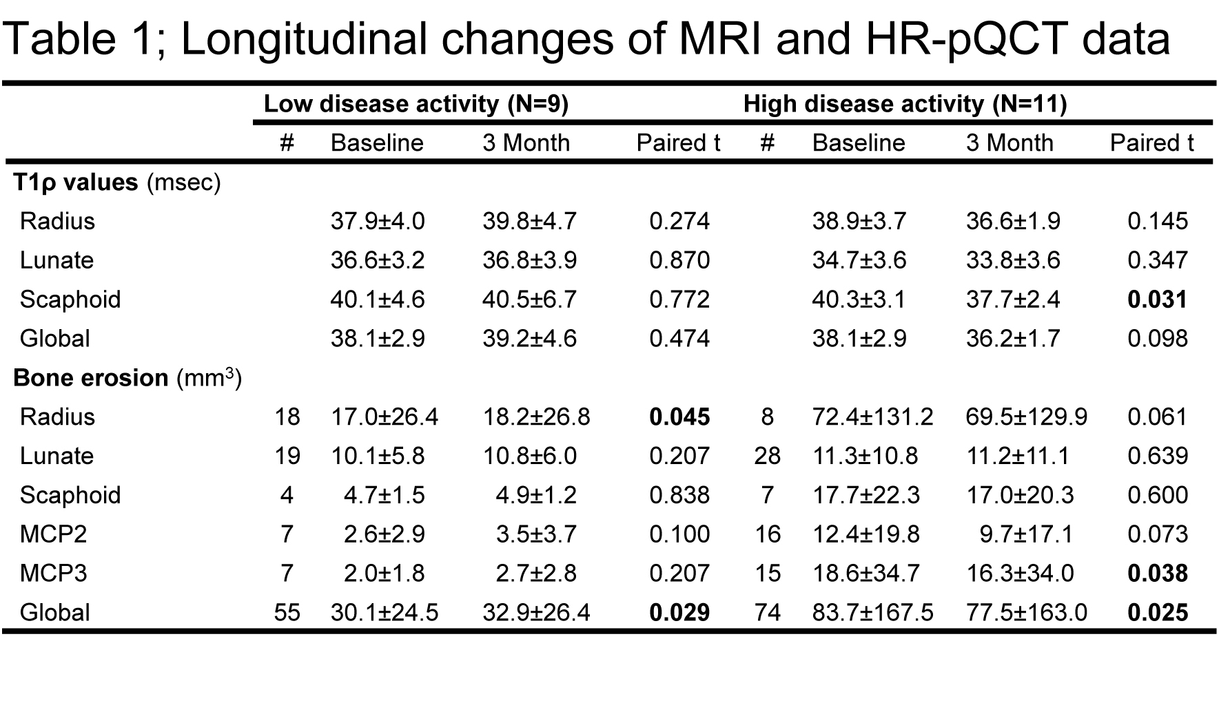
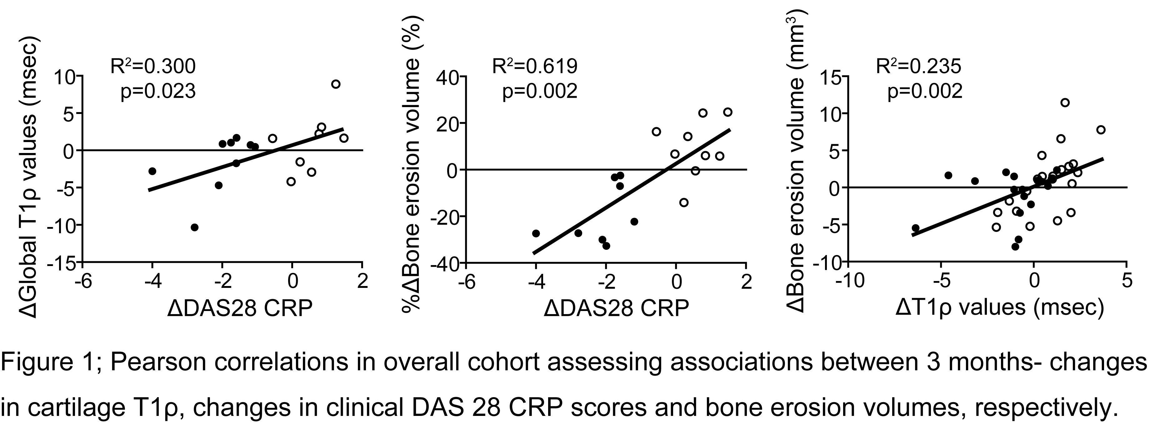
Results: Anti-TNFƒ¿ therapy in the high-DAS group resulted in a significant decrease of DAS28 CRP score (p<0.001), which was accompanied by decreases in mean T1ƒÏ values (reached significance in scaphoid T1rho, P=0.031) and erosion volumes (reached significance in MCP3 and global) at all measurement sites (Table.1). The low DAS group in contrast, displayed an increasing trend in T1rho values and erosion volumes (reached significance in radius and global) despite low disease activity (Table.1) Changes in T1rho values and bone erosion volume were significantly positively correlated, and both were significantly correlated with changes in DAS28 CRP score (Figure 1).

Conclusion: Anti-TNFƒ¿ therapy seems to simultaneously prevent bone and cartilage from further destruction and might even partially repair preexisting cartilage deterioration and bone erosions within first 3 months of therapy as indicated by our observed decrease in T1rho values and erosion volumes in our cohort. Our observation that early changes in cartilage T1rho values are associated with changes in clinical disease activity and erosion volume, recommends a more extensive investigation of T1rho as a predictor of disease progression. Our data illustrate the powerful potential of multimodal imaging with MRI and HR-pQCT to evaluate early response to treatment in RA.


Disclosure: T. Shimizu, None; K. Mamoto, None; U. Heilmeier, None; M. Tanaka, None; A. J. Burghardt, None; T. Link, None; J. Graf, None; J. B. Imboden Jr., None; X. Li, None.

To cite this abstract in AMA style:

Shimizu T, Mamoto K, Heilmeier U, Tanaka M, Burghardt AJ, Link T, Graf J, Imboden JB Jr., Li X. Changes in Cartilage Matrix Measured By MR T1ρ Are Correlated with Changes in Bone Erosion Volume Measured By HR-pQCT Three Months after MTX and Anti-TNF Treatment in Patients with Rheumatoid Arthritis: A Multi-Modality Imaging Study [abstract]. Arthritis Rheumatol. 2017; 69 (suppl 10). https://acrabstracts.org/abstract/changes-in-cartilage-matrix-measured-by-mr-t1%cf%81-are-correlated-with-changes-in-bone-erosion-volume-measured-by-hr-pqct-three-months-after-mtx-and-anti-tnf-treatment-in-patients-with-rheumatoid-art/. Accessed .
  • Tweet
  • Click to email a link to a friend (Opens in new window) Email
  • Click to print (Opens in new window) Print

« Back to 2017 ACR/ARHP Annual Meeting

ACR Meeting Abstracts - https://acrabstracts.org/abstract/changes-in-cartilage-matrix-measured-by-mr-t1%cf%81-are-correlated-with-changes-in-bone-erosion-volume-measured-by-hr-pqct-three-months-after-mtx-and-anti-tnf-treatment-in-patients-with-rheumatoid-art/

Advanced Search

Your Favorites

You can save and print a list of your favorite abstracts during your browser session by clicking the “Favorite” button at the bottom of any abstract. View your favorites »

All abstracts accepted to ACR Convergence are under media embargo once the ACR has notified presenters of their abstract’s acceptance. They may be presented at other meetings or published as manuscripts after this time but should not be discussed in non-scholarly venues or outlets. The following embargo policies are strictly enforced by the ACR.

Accepted abstracts are made available to the public online in advance of the meeting and are published in a special online supplement of our scientific journal, Arthritis & Rheumatology. Information contained in those abstracts may not be released until the abstracts appear online. In an exception to the media embargo, academic institutions, private organizations, and companies with products whose value may be influenced by information contained in an abstract may issue a press release to coincide with the availability of an ACR abstract on the ACR website. However, the ACR continues to require that information that goes beyond that contained in the abstract (e.g., discussion of the abstract done as part of editorial news coverage) is under media embargo until 10:00 AM ET on November 14, 2024. Journalists with access to embargoed information cannot release articles or editorial news coverage before this time. Editorial news coverage is considered original articles/videos developed by employed journalists to report facts, commentary, and subject matter expert quotes in a narrative form using a variety of sources (e.g., research, announcements, press releases, events, etc.).

Violation of this policy may result in the abstract being withdrawn from the meeting and other measures deemed appropriate. Authors are responsible for notifying colleagues, institutions, communications firms, and all other stakeholders related to the development or promotion of the abstract about this policy. If you have questions about the ACR abstract embargo policy, please contact ACR abstracts staff at [email protected].

Wiley

  • Online Journal
  • Privacy Policy
  • Permissions Policies
  • Cookie Preferences

© Copyright 2025 American College of Rheumatology