Session Information
Session Type: ACR Poster Session C
Session Time: 9:00AM-11:00AM
Background/Purpose: MR-based T1rho relaxation time mapping allows non-invasive quantification of early cartilage deterioration, while high-resolution peripheral quantitative computed tomography (HR-pQCT) can detect early changes in bone erosion volume. It is unknown whether changes of cartilage T1rho values are associated with concomitant bone erosion changes and reflect treatment response. Therefore, we used 3T MRI and HR-pQCT to investigate the changes of bone and cartilage damage in RA patients receiving methotrexate (MTX) and anti-tumor necrosis factor alpha (TNFa) therapy.
Methods: Twenty RA patients receiving MTX treatment were recruited into either a low DAS group (n=9, DAS28≤3.2) or a high DAS group (n=11, DAS28>3.2). At baseline, the high DAS group initiated supplemental anti-TNFa treatment in addition to ongoing MTX. All patients underwent MRI wrist scans and HR-pQCT scans of the MCP and wrist at BL and after 3 months (3M). DAS28 CRP was assessed at BL and 3M. Lunar, scaphoid and radius and global cartilage T1rho values were measured. HR-pQCT-derived erosion volume at lunate, radius, scaphoid, MCP2 and MCP3 were semi-quantitatively measured. Longitudinal changes and associations were evaluated either via paired t-test or Pearsonfs correlations.
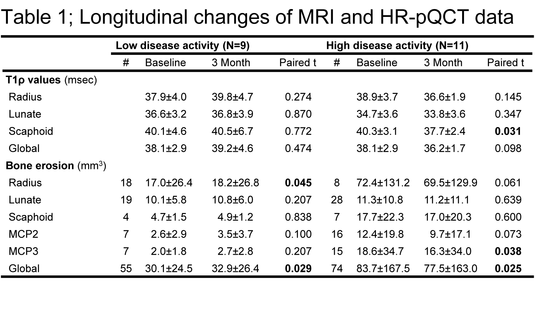
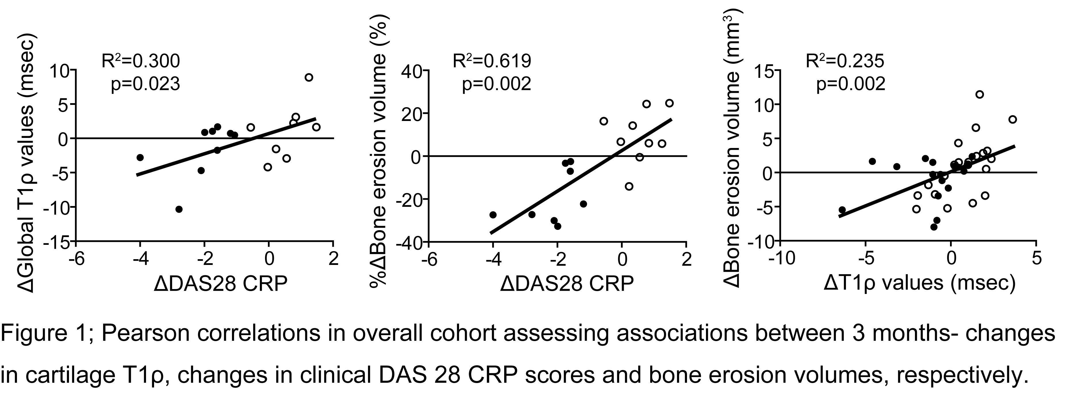
Results: Anti-TNFƒ¿ therapy in the high-DAS group resulted in a significant decrease of DAS28 CRP score (p<0.001), which was accompanied by decreases in mean T1ƒÏ values (reached significance in scaphoid T1rho, P=0.031) and erosion volumes (reached significance in MCP3 and global) at all measurement sites (Table.1). The low DAS group in contrast, displayed an increasing trend in T1rho values and erosion volumes (reached significance in radius and global) despite low disease activity (Table.1) Changes in T1rho values and bone erosion volume were significantly positively correlated, and both were significantly correlated with changes in DAS28 CRP score (Figure 1).
Conclusion: Anti-TNFƒ¿ therapy seems to simultaneously prevent bone and cartilage from further destruction and might even partially repair preexisting cartilage deterioration and bone erosions within first 3 months of therapy as indicated by our observed decrease in T1rho values and erosion volumes in our cohort. Our observation that early changes in cartilage T1rho values are associated with changes in clinical disease activity and erosion volume, recommends a more extensive investigation of T1rho as a predictor of disease progression. Our data illustrate the powerful potential of multimodal imaging with MRI and HR-pQCT to evaluate early response to treatment in RA.
To cite this abstract in AMA style:
Shimizu T, Mamoto K, Heilmeier U, Tanaka M, Burghardt AJ, Link T, Graf J, Imboden JB Jr., Li X. Changes in Cartilage Matrix Measured By MR T1ρ Are Correlated with Changes in Bone Erosion Volume Measured By HR-pQCT Three Months after MTX and Anti-TNF Treatment in Patients with Rheumatoid Arthritis: A Multi-Modality Imaging Study [abstract]. Arthritis Rheumatol. 2017; 69 (suppl 10). https://acrabstracts.org/abstract/changes-in-cartilage-matrix-measured-by-mr-t1%cf%81-are-correlated-with-changes-in-bone-erosion-volume-measured-by-hr-pqct-three-months-after-mtx-and-anti-tnf-treatment-in-patients-with-rheumatoid-art/. Accessed .« Back to 2017 ACR/ARHP Annual Meeting
ACR Meeting Abstracts - https://acrabstracts.org/abstract/changes-in-cartilage-matrix-measured-by-mr-t1%cf%81-are-correlated-with-changes-in-bone-erosion-volume-measured-by-hr-pqct-three-months-after-mtx-and-anti-tnf-treatment-in-patients-with-rheumatoid-art/


