Session Information
Date: Sunday, November 8, 2015
Title: Systemic Lupus Erythematosus - Human Etiology and Pathogenesis Poster I
Session Type: ACR Poster Session A
Session Time: 9:00AM-11:00AM
Background/Purpose:
Discoid
lupus erythematosus (DLE) is the most common form of chronic cutaneous lupus. Lesional
skin of DLE patients contains macrophages that may transition from a
pro-inflammatory M1 subtype to an anti-inflammatory M2 subtype, and vice
versa. To test our hypothesis that M1 macrophages would be increased in DLE
skin, we examined macrophage features in DLE and normal skin using
histochemical and gene expression approaches.
Methods:
DLE
lesional and normal control skin samples were analyzed for CD163 expression by
immunohistochemistry. Gene expression of RNA from both skin groups was compared
by microarrays and quantitative RT-PCR. Double immunofluorescence studies were
performed to characterize protein expression of CD163+ macrophages.
Results:
CD163+
macrophages were increased near the epidermal-dermal junction and perivascular
areas in DLE lesional skin compared with normal skin. Gene set enrichment
analysis comparing differentially expressed genes in DLE and normal skin with
previously published gene sets associated with M1 and M2 macrophages [1] showed
strong overlap between up-regulated genes in DLE skin and M1 macrophages
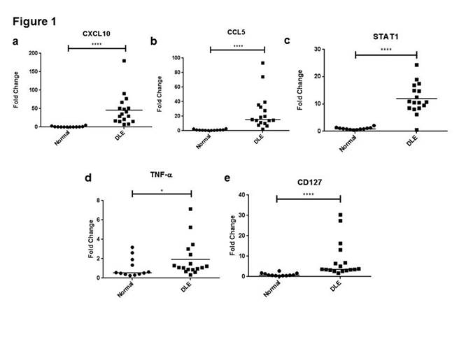
(p=0.02) (Table 1). Quantitative RT-PCR showed that several M1
macrophage-associated genes (e.g. CXCL10 (47.2 fold change (FC), CCL5 (25.9
FC), STAT1 (11.96 FC), TNF-α (1.92 FC), CD127 (8.01 FC), p<0.05) had
amplified mRNA levels in DLE skin (Figure 1a-e). However, double
immunofluorescence studies of CD163+ macrophages revealed co-expression
of M1 (CXCL10, TNF-α, CD127), and M2 (CD209, TGF-β)
macrophage-related proteins in DLE skin.
Conclusion:
The
variation of macrophage subtypes in DLE skin may reflect their propensity to
switch polarization and possible transition from a M1-dominant acute phase to
an M2-dominant chronic phase, though this requires verification. Macrophage
plasticity makes these cells ideal targets for future therapies.
References:
1.
Fuentes-Duculan J, Suarez-Farinas M, Zaba LC, Nograles KE, Pierson KC, Mitsui H
et al. A subpopulation of CD163-positive macrophages is classically
activated in psoriasis. J Invest Dermatol 130 (2010) 2412-22.
Table
1. Gene set enrichment analysis of differentially expressed genes in DLE
lesional and normal skin.
|
Name |
Size |
ES |
NES |
p-value |
FDR q-value |
|
IFN-γ-stimulated (M1) macrophages up-regulated |
113 |
0.92 |
1.25 |
0.02 |
0.02 |
|
IFN-γ stimulated (M1) macrophages down-regulated |
16 |
-0.55 |
-1.24 |
0.25 |
0.25 |
|
IL-4 stimulated (M2) macrophages up-regulated |
26 |
0.39 |
0.97 |
0.49 |
0.49 |
|
IL-4 stimulated (M2) macrophages down-regulated |
1 |
0.95 |
1.11 |
0.27 |
0.27 |
Abbreviations:
DLE – discoid lupus erythematosus, ES – enrichment score, FDR – false discovery
rate, IFN – interferon, IL – interleukin, NES – normalized enrichment score
To cite this abstract in AMA style:
Chong B, Tseng LC, Hosler G, Teske N, Zhang S, Karp DR, Olsen NJ, Mohan C. CD163+ Macrophages Display Mixed Polarizations in Discoid Lupus Skin [abstract]. Arthritis Rheumatol. 2015; 67 (suppl 10). https://acrabstracts.org/abstract/cd163-macrophages-display-mixed-polarizations-in-discoid-lupus-skin/. Accessed .« Back to 2015 ACR/ARHP Annual Meeting
ACR Meeting Abstracts - https://acrabstracts.org/abstract/cd163-macrophages-display-mixed-polarizations-in-discoid-lupus-skin/

