ACR Meeting Abstracts

ACR Meeting Abstracts

  • Meetings
    • ACR Convergence 2024
    • ACR Convergence 2023
    • 2023 ACR/ARP PRSYM
    • ACR Convergence 2022
    • ACR Convergence 2021
    • ACR Convergence 2020
    • 2020 ACR/ARP PRSYM
    • 2019 ACR/ARP Annual Meeting
    • 2018-2009 Meetings
    • Download Abstracts
  • Keyword Index
  • Advanced Search
  • Your Favorites
    • Favorites
    • Login
    • View and print all favorites
    • Clear all your favorites
  • ACR Meetings

Abstract Number: 1299

Can the 3D-Modified Dixon Methods Replace the Conventional Sacroiliac Joint Magnetic Resonance Imaging?

Yoonah Song1, Seunghun Lee1 and Tae-Hwan Kim2, 1Department of Radiology, Hanyang University College of Medicine, Seoul Hospital, Seoul, South Korea, 2Department of Rheumatology, Department of Rheumatology, Hanyang University Hospital for Rheumatic Diseases, Seoul, South Korea

Meeting: 2016 ACR/ARHP Annual Meeting

Date of first publication: September 28, 2016

Keywords: magnetic resonance imaging (MRI) and spondylarthritis

  • Tweet
  • Click to email a link to a friend (Opens in new window) Email
  • Click to print (Opens in new window) Print
Session Information

Date: Monday, November 14, 2016

Title: Imaging of Rheumatic Diseases - Poster II: XR/CT/PET/MRI

Session Type: ACR Poster Session B

Session Time: 9:00AM-11:00AM

Can the 3D-modified Dixon

Methods Replace the Conventional Sacroiliac Joint Magnetic Resonance Imaging?  

ABSTRACT

Background/Purpose: The purpose of our study was to assess the diagnostic value of two-point Dixon method compared with conventional magnetic resonance imaging (MRI) including T1-weighted, T2-weighted, fat saturated (FS) T1-weighted, FS T2-weighted, and short tau inversion recovery (STIR) in diagnosing axial spondyloarthritis (SpA). Methods: A total of 67 patients with low back pain and suspected axial SpA who underwent sacroiliac joint MRI in our institution from September 2014 to October 2015 were retrospectively evaluated. Each with two data sets (modified Dixon images, conventional MRI), were independently reviewed by two radiologists as follows: 1) grading of both sacroiliac joints; 2) bone marrow edema; 3) erosion; 4) periarticular fat deposition; 5) ankylosis. Bone marrow edema was evaluated on FS T2-weighted, and STIR images. Structural change and fat deposition were evaluated on T1-weighted image. Erosion was evaluated on T1-weighted and FS T1-weighted images. All variables were determined in a thin slice 3D-Dixon images. Sensitivity, specificity, and intra- and interobserver reliabilities were calculated.

Results: Patients had a mean age of 31.7 ¡¾ 11.1 years and male were 44 (65.7%). 44 of 67 patients (65.7%) had positive HLA-B27. 62 patients were diagnosed with axial SpA based on laboratory and imaging information as well as the Assessment of Spondyloarthritis International Society (ASAS) criteria. One patient was diagnosed with osteitis condensans ilii. Four patients were diagnosed with non-specific mechanical back pain. Sensitivities and specificities of Dixon method and conventional MRI are 97.4%, 14.3% and 94.6%, 10.7%. Both Dixon method and conventional MRI had a good to excellent intraobserver (mean intraclass correlation coefficient [ICC] = 0.680-0.953, 0.886-1.000) and interobserver agreement (mean ICC = 0.860-0.986, 0.792-0.953).

Conclusion: 3D-modified Dixon method might be another practical novel imaging technique as conventional MRI. The Dixon method is more intuitive than conventional MRI to evaluate sacroiliac joint, moreover, it takes less time.

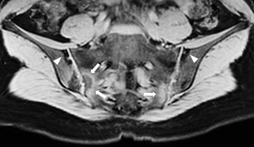
Figure 1. Bilateral sacroiliitis (grade 3) with periarticular fat deposition (arrow head), and edema (arrow) showed in 3D-Dixon methods.


Disclosure: Y. Song, None; S. Lee, None; T. H. Kim, None.

To cite this abstract in AMA style:

Song Y, Lee S, Kim TH. Can the 3D-Modified Dixon Methods Replace the Conventional Sacroiliac Joint Magnetic Resonance Imaging? [abstract]. Arthritis Rheumatol. 2016; 68 (suppl 10). https://acrabstracts.org/abstract/can-the-3d-modified-dixon-methods-replace-the-conventional-sacroiliac-joint-magnetic-resonance-imaging/. Accessed .
  • Tweet
  • Click to email a link to a friend (Opens in new window) Email
  • Click to print (Opens in new window) Print

« Back to 2016 ACR/ARHP Annual Meeting

ACR Meeting Abstracts - https://acrabstracts.org/abstract/can-the-3d-modified-dixon-methods-replace-the-conventional-sacroiliac-joint-magnetic-resonance-imaging/

Advanced Search

Your Favorites

You can save and print a list of your favorite abstracts during your browser session by clicking the “Favorite” button at the bottom of any abstract. View your favorites »

All abstracts accepted to ACR Convergence are under media embargo once the ACR has notified presenters of their abstract’s acceptance. They may be presented at other meetings or published as manuscripts after this time but should not be discussed in non-scholarly venues or outlets. The following embargo policies are strictly enforced by the ACR.

Accepted abstracts are made available to the public online in advance of the meeting and are published in a special online supplement of our scientific journal, Arthritis & Rheumatology. Information contained in those abstracts may not be released until the abstracts appear online. In an exception to the media embargo, academic institutions, private organizations, and companies with products whose value may be influenced by information contained in an abstract may issue a press release to coincide with the availability of an ACR abstract on the ACR website. However, the ACR continues to require that information that goes beyond that contained in the abstract (e.g., discussion of the abstract done as part of editorial news coverage) is under media embargo until 10:00 AM ET on November 14, 2024. Journalists with access to embargoed information cannot release articles or editorial news coverage before this time. Editorial news coverage is considered original articles/videos developed by employed journalists to report facts, commentary, and subject matter expert quotes in a narrative form using a variety of sources (e.g., research, announcements, press releases, events, etc.).

Violation of this policy may result in the abstract being withdrawn from the meeting and other measures deemed appropriate. Authors are responsible for notifying colleagues, institutions, communications firms, and all other stakeholders related to the development or promotion of the abstract about this policy. If you have questions about the ACR abstract embargo policy, please contact ACR abstracts staff at [email protected].

Wiley

  • Online Journal
  • Privacy Policy
  • Permissions Policies
  • Cookie Preferences

© Copyright 2025 American College of Rheumatology