ACR Meeting Abstracts

ACR Meeting Abstracts

  • Meetings
    • ACR Convergence 2024
    • ACR Convergence 2023
    • 2023 ACR/ARP PRSYM
    • ACR Convergence 2022
    • ACR Convergence 2021
    • ACR Convergence 2020
    • 2020 ACR/ARP PRSYM
    • 2019 ACR/ARP Annual Meeting
    • 2018-2009 Meetings
    • Download Abstracts
  • Keyword Index
  • Advanced Search
  • Your Favorites
    • Favorites
    • Login
    • View and print all favorites
    • Clear all your favorites
  • ACR Meetings

Abstract Number: 1272

Can Histologically Defined Peri-Articular Vascular Channels be Identified on High-Resolution Computed Tomography? – a Study in Cadaveric Finger Joints

A. Scharmga1, K. K. Keller2, M. Peters1, A. van Tubergen1, J. van den Bergh3, B. van Rietbergen4, R. Weijers5, D. Loeffen5, E-M. Hauge2 and P. Geusens6, 1Department of Internal Medicine, Rheumatology, Maastricht University Medical Center, Maastricht, Netherlands, 2Rheumatology, Department of Rheumatology, Aarhus University Hospital, Aarhus, Denmark, 3Internal Medicine, Viecuri Medical Center, Venlo, Netherlands, 4Biomedical Engineering, Eindhoven University of Technology, Eindhoven, Netherlands, 5Department of Radiology, Maastricht University Medical Center, Maastricht, Netherlands, 6Internal Medicine/Rheumatology, Maastricht University Medical Center and Hasselt University, Biomedical Research Institute, Diepenbeek, Belgium

Meeting: 2016 ACR/ARHP Annual Meeting

Date of first publication: September 28, 2016

Keywords: Imaging and joint damage

  • Tweet
  • Click to email a link to a friend (Opens in new window) Email
  • Click to print (Opens in new window) Print
Session Information

Date: Monday, November 14, 2016

Title: Imaging of Rheumatic Diseases - Poster II: XR/CT/PET/MRI

Session Type: ACR Poster Session B

Session Time: 9:00AM-11:00AM

Background/Purpose: Several studies have indicated that High Resolution peripheral CT (HR-pQCT) scanning is more sensitive than radiography in detecting cortical interruptions in destructive joint diseases like rheumatoid arthritis (RA)[1-3]. Cortical interruptions are also seen in healthy controls, but the exact nature of these interruptions is not known, and might represent vascular channels (VCs). No previous study has compared histology to HR-pQCT images in finger joints. We hypothesized that VCs seen on histology can also be detected by HR-pQCT imaging.

Methods: Based on HR-pQCT, 4 regions in 3 metacarpophalangeal (MCP) joints from female cadavers with an unknown medical history (mean age 84.7, SD 5.5 years) were selected. These regions were extracted, embedded undecalcified in methylmetacrylate and histologically sectioned (thickness 14μm) parallel to the axial plane. Every second section (n=478) was stained with Goldner Trichrome. VCs were identified as a cortical interruption in one histological section, which contained one or more vessels. Histological sections were matched visually to corresponding axial HR-pQCT images. Per match, it was described if a cortical interruption was observed on HR-pQCT and if this was a VC based on an interruption with a parallel structure present on 2 consecutive slices in 2 planes (axial, and/or sagittal/coronal).

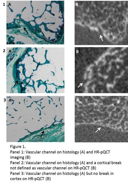
Results: A total of 52 different VCs were identified on histology. All could be matched to a corresponding axial HR-pQCT image. Twenty histologically defined VCs would be defined as VC on HR-pQCT based on the parallel structure, although only 5 were present on 2 consecutive slices and 15 on less than 2 consecutive slices. Twenty-seven interruptions were observed on HR-pQCT, but did not fulfill the definition of a VC. In 5 cases no interruptions could be observed on HR-pQCT. Figure 1 demonstrates 3 examples of histology and matching HR-pQCT images.

Conclusion: VCs were frequently present in MCP joints. Only a minority of histologically defined VCs is interpreted as VC using a pre-specified definition on HR-pQCT images. Small histological VCs were often identified as an interruption but rarely considered a VC on HR-pQCT. Therefore, additional criteria in order to categorize a VC as such on HR-pQCT are warranted.  

References.1.Stach 2010 2.Srikhum 2013.3.Fouque-Aubert 2010


Disclosure: A. Scharmga, None; K. K. Keller, None; M. Peters, None; A. van Tubergen, None; J. van den Bergh, None; B. van Rietbergen, Scanco Medical A.G., 5; R. Weijers, None; D. Loeffen, None; E. M. Hauge, None; P. Geusens, None.

To cite this abstract in AMA style:

Scharmga A, Keller KK, Peters M, van Tubergen A, van den Bergh J, van Rietbergen B, Weijers R, Loeffen D, Hauge EM, Geusens P. Can Histologically Defined Peri-Articular Vascular Channels be Identified on High-Resolution Computed Tomography? – a Study in Cadaveric Finger Joints [abstract]. Arthritis Rheumatol. 2016; 68 (suppl 10). https://acrabstracts.org/abstract/can-histologically-defined-peri-articular-vascular-channels-be-identified-on-high-resolution-computed-tomography-a-study-in-cadaveric-finger-joints/. Accessed .
  • Tweet
  • Click to email a link to a friend (Opens in new window) Email
  • Click to print (Opens in new window) Print

« Back to 2016 ACR/ARHP Annual Meeting

ACR Meeting Abstracts - https://acrabstracts.org/abstract/can-histologically-defined-peri-articular-vascular-channels-be-identified-on-high-resolution-computed-tomography-a-study-in-cadaveric-finger-joints/

Advanced Search

Your Favorites

You can save and print a list of your favorite abstracts during your browser session by clicking the “Favorite” button at the bottom of any abstract. View your favorites »

All abstracts accepted to ACR Convergence are under media embargo once the ACR has notified presenters of their abstract’s acceptance. They may be presented at other meetings or published as manuscripts after this time but should not be discussed in non-scholarly venues or outlets. The following embargo policies are strictly enforced by the ACR.

Accepted abstracts are made available to the public online in advance of the meeting and are published in a special online supplement of our scientific journal, Arthritis & Rheumatology. Information contained in those abstracts may not be released until the abstracts appear online. In an exception to the media embargo, academic institutions, private organizations, and companies with products whose value may be influenced by information contained in an abstract may issue a press release to coincide with the availability of an ACR abstract on the ACR website. However, the ACR continues to require that information that goes beyond that contained in the abstract (e.g., discussion of the abstract done as part of editorial news coverage) is under media embargo until 10:00 AM ET on November 14, 2024. Journalists with access to embargoed information cannot release articles or editorial news coverage before this time. Editorial news coverage is considered original articles/videos developed by employed journalists to report facts, commentary, and subject matter expert quotes in a narrative form using a variety of sources (e.g., research, announcements, press releases, events, etc.).

Violation of this policy may result in the abstract being withdrawn from the meeting and other measures deemed appropriate. Authors are responsible for notifying colleagues, institutions, communications firms, and all other stakeholders related to the development or promotion of the abstract about this policy. If you have questions about the ACR abstract embargo policy, please contact ACR abstracts staff at [email protected].

Wiley

  • Online Journal
  • Privacy Policy
  • Permissions Policies
  • Cookie Preferences

© Copyright 2025 American College of Rheumatology