Session Information
Date: Sunday, October 21, 2018
Title: Rheumatoid Arthritis – Diagnosis, Manifestations, and Outcomes Poster I: Comorbidities
Session Type: ACR Poster Session A
Session Time: 9:00AM-11:00AM
Background/Purpose: Rheumatoid arthritis (RA) is described as an independent risk factor for osteoporosis and is included in models, including the FRAX™ tool which uses bone mineral density (BMD) as well as demographics to predict fracture risk. Most dual X-ray absorptiometry (DEXA) scans only measure the non-dominant hip. Published data are from existing datasets that are X-sectional and the influence of factors on progression of BMD loss in different sites has not be studied. We aimed to test the hypothesis that bone loss over time is more pronounced in patients with RA, compared to those without.
Methods: We used data from patients referred for DEXA in the North West of England from June 2004 and October 2016. Patients have their FRAX™ risk factors assessed and their BMD measured in both femoral necks, shaft, trochanter and Wards triangle in addition to L1-L4 in the lumbar spine (LS). Patients attending for multiple scans in this period without a diagnosis of RA were used as a comparator group. A longitudinal mixed-effects model was fitted, with BMD at each site as the exposure, adjusting for all risk factors for OP in addition to fitting interaction terms for bisphosphonate use in addition to calcium/vitamin D use.
Results:
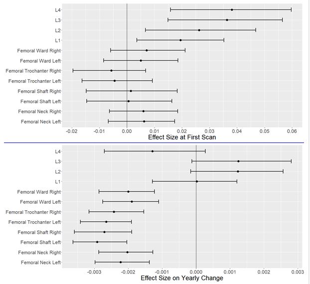
6941 patients were included, 6169 (88.9%) were females. Of whom 1270 (20.5%) were menopausal. Mean age was 63 years (SD 11.3 years) and 749 patients with RA (named the RA group) had multiple scans and were compared to the 6192 without an RA diagnosis. There are an average of 2.35 scans per patient (SD 0.64 scans),an average of 4.26 years apart (SD 1.88 years). Patients in the RA group had significantly higher average BMD in the lumber spine than those in the non-RA group. There was no significant difference in the bone mineral density of the femur between the RA and non-RA groups. Conversely patients in the RA group lost BMD faster in the femur than the non-RA group, but BMD loss in the lumbar spine was not significantly different between groups.
Figure 1. Showing the results of the longitudinal analysis for average BMD (top) and bone loss over time (bottom)
Conclusion: The average BMD in the lumbar spine is higher in the RA group than in the non-RA group, but bone loss appears to be increased in the femur at all sites of measurement. The most likely explanation is increasing OA in the lumbar spine causing slower loss of BMD. This is clinically significant because bone loss in the hip is used as a predictor for fracture and the rapid bone loss seen would necessitate reducing the interval between scans in this group of patients. Limitations of the study include lack of information about length of disease and specific treatments history for the RA patients.
To cite this abstract in AMA style:
Tribbick R, Massarotti M, Kerns JG, Dondelinger F, Bukhari M. Bone Loss in Different Sites over Time in Patients with Rheumatoid Arthritis [abstract]. Arthritis Rheumatol. 2018; 70 (suppl 9). https://acrabstracts.org/abstract/bone-loss-in-different-sites-over-time-in-patients-with-rheumatoid-arthritis/. Accessed .« Back to 2018 ACR/ARHP Annual Meeting
ACR Meeting Abstracts - https://acrabstracts.org/abstract/bone-loss-in-different-sites-over-time-in-patients-with-rheumatoid-arthritis/

