Session Information
Session Type: ACR Poster Session A
Session Time: 9:00AM-11:00AM
Background/Purpose: Giant cell arteritis (GCA) is the most common type of systemic vasculitis. Currently, two forms of GCA are described: a cranial(C)-GCA (temporal arteritis) and a systemic, large-vessel (LV)-GCA. Late complications of LV-GCA are aortic aneurysms or aortic rupture. Based on the analysis of temporal artery tissue, GCA is postulated to start at the adventitial site and to be T cell-mediated. In the temporal artery infiltrates, T cells clearly outnumber B cells. However, our report on a disturbed homeostasis of B cells in newly diagnosed C-GCA patients shows evidence for a putative role of B cells in GCA.1 Recently, the presence of B cells organized in artery tertiary lymphoid organs (ATLOs) has been described in C-GCA.2 The immunopathological role of B cells in both forms of GCA is underexplored and it is unknown whether B cells are present in the vessel wall of patients with LV-GCA. The objective of this study was to assess the presence and organization of B cells in the aorta of patients with LV-GCA.
Methods: Aorta tissue samples of 11 histologically-proven LV-GCA patients who underwent surgery due to an aortic aneurysm were studied by immunohistochemistry. Staining was performed with antibodies against CD20 (B cells), CD3 (T cells), CD21 (follicular dendritic cells (FDC)), PNAd (high endothelial venules (HEV)), bcl6 (germinal centers), and CD138 (plasma cells). None of the patients was receiving immunosuppressive treatment at the time of surgery. For comparison 22 aorta samples from age- and sex-matched atherosclerosis patients with an aortic aneurysm were included.
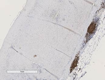
Results: Aorta tissues of LV-GCA patients showed massive infiltration of B cells (see fig. 1). The infiltrating B cells were mainly found in the adventitia and were organized into high density B cell areas. In contrast to the temporal artery, B cells outnumbered T cells in the aorta. Besides the presence of T cells, FDC, germinal centers, plasma cells, and HEV were documented at the areas of B cell infiltrates, which are typical for organized ATLOs.
Conclusion: Aorta tissues from patients with histologically-proven LV-GCA showed massive and organized B cell infiltrates mostly located in the adventitia. The mere presence of B cells at the site of inflammation prompts further investigation into the role of B cells in the pathogenesis of GCA.
1. van der Geest KSM, Abdulahad WH, Chalan P, et al. Disturbed B cell homeostasis in newly diagnosed giant cell arteritis and polymyalgia rheumatica. Art Rheumatol. 2014;66(7):1927-1938.
2. Ciccia F, Rizzo A, Maugeri R, et al. Ectopic expression of CXCL13, BAFF, APRIL and LT-β is associated with artery tertiary lymphoid organs in giant cell arteritis. Ann Rheum Dis. 2016;0:1-9.
Fig. 1. Aorta from a LV-GCA patient with organized high density
CD20+ B cells areas in the adventitia (DAB/brown).
To cite this abstract in AMA style:
Graver JC, Sandovici M, Abdulahad WH, Haacke EA, Boots AMH, Brouwer E. B Cells in Giant Cell Arteritis: a Novel Target for Treatment? [abstract]. Arthritis Rheumatol. 2017; 69 (suppl 10). https://acrabstracts.org/abstract/b-cells-in-giant-cell-arteritis-a-novel-target-for-treatment/. Accessed .« Back to 2017 ACR/ARHP Annual Meeting
ACR Meeting Abstracts - https://acrabstracts.org/abstract/b-cells-in-giant-cell-arteritis-a-novel-target-for-treatment/

