ACR Meeting Abstracts

ACR Meeting Abstracts

  • Meetings
    • ACR Convergence 2024
    • ACR Convergence 2023
    • 2023 ACR/ARP PRSYM
    • ACR Convergence 2022
    • ACR Convergence 2021
    • ACR Convergence 2020
    • 2020 ACR/ARP PRSYM
    • 2019 ACR/ARP Annual Meeting
    • 2018-2009 Meetings
    • Download Abstracts
  • Keyword Index
  • Advanced Search
  • Your Favorites
    • Favorites
    • Login
    • View and print all favorites
    • Clear all your favorites
  • ACR Meetings

Abstract Number: 1075

Automated Multiparameter Microscopy and Image Analysis: Next Generation Synovial Tissue Histology

William Murray-Brown1, Helen Weedon1, Amanda Aloia2, Susan Lester3, Susanna Proudman4, Malcolm D. Smith5, Jenny Walker1 and Mihir D Wechalekar1,3, 1Rheumatology, Flinders University, Adelaide, Australia, 2Cellular Imaging, Flinders University, Adelaide, Australia, 3Royal Adelaide Hospital, Adelaide, Australia, 4Royal Adelaide Hospital, Rheumatology Unit and University of Adelaide, Discipline of Medicine, Adelaide, Australia, 5Rheumotology, Flinders University, Adelaide, Australia

Meeting: 2018 ACR/ARHP Annual Meeting

Keywords: imaging techniques, rheumatoid arthritis (RA) and synovial cells, synovial fluid

  • Tweet
  • Click to email a link to a friend (Opens in new window) Email
  • Click to print (Opens in new window) Print
Session Information

Date: Monday, October 22, 2018

Title: Rheumatoid Arthritis – Etiology and Pathogenesis Poster II

Session Type: ACR Poster Session B

Session Time: 9:00AM-11:00AM

Background/Purpose: Currently, histological assessment of synovial tissue (ST) in Rheumatoid Arthritis (RA) is hampered by 3 main factors: 1) tissue is a finite resource, 2) sequential sections of single conventional immunohistochemistry stains do not adequately preserve architectural and co-staining data, and, 3) image analysis and quantification is open to interpretation and human error. Furthermore,in RA, growing evidence implicates heterogeneous histopathological phenotypes with different disease outcomes and treatment responses, critical, as 30-40% of patients do not respond to current therapies and long-term drug-remission is rare, reflecting differential recruitment of inflammatory pathways and cell types. Thus, there is an unmet need for tools that can perform automated multi-parameter cellular identification and phenotype analysis on tissue sections, enabling translational research to assist is the development of personalised medicine.

Methods: Sequential 4μm thick RA ST sections were prepared for Opal multispectral imaging. In brief, for Opal; primary antibodies identifying memory T (CD45RO), Β (CD20) and macrophage cell (CD68) are stained in turn, each followed by specific Opal reactive fluorophores. Primary antibody stripping prepares the section for the next primary antibody ready to repeat the process. Once all Opal targets have been developed, DAPI nuclear staining is performed prior to whole slide scanning (Figure 1A) and high-resolution (x20 magnification) image acquisition for CD45RO (green), CD20 (red), CD68 (cyan) and DAPI nuclear stain (blue) (Figure 1B) on the Perkin Elmer Vectra 3.0 automated imaging system.

Results: The image analysis and machine learning software package, inForm® was used to segment individual cells (Figure 1C), identify and quantify cellular phenotype (Figure 1D&E) and distinguish regions with “follicular” and “diffuse” staining patterns (Figure 1F&G).

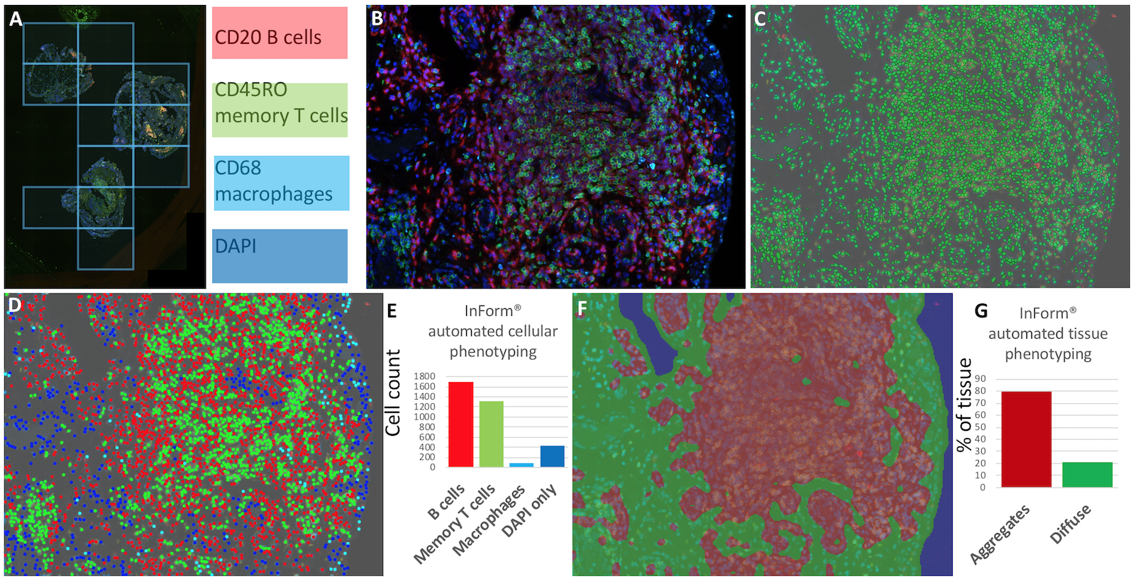
Figure 1. Automated multiparameter imaging and analysis of RA ST. A, whole slide scanning. Β, high-resolution multiparameter microscopy for CD20 (red), CD45RO (green) and CD68 (cyan). C, cell segmentation map. D, cellular phenotyping and E, automated cellular counting. F, tissue phenotyping and G, automated phenotype assessment.

Conclusion: Due to the recent surge in interest in ST biology, in particular in clinical trials investigating biologic therapies, there is a need for effective, reproducible and high-throughout technologies for immune-histological examination. Thus, a tool such as Opal can be utilised for whole slide scanning and automated analysis of ST infiltrates and tissue phenotype analysis. ST phenotype is thought to influence treatment outcomes in patients, therefore, comprehensive analysis of ST histology will result in more personalised and stratified medicine regimes for patients.


Disclosure: W. Murray-Brown, None; H. Weedon, None; A. Aloia, None; S. Lester, None; S. Proudman, None; M. D. Smith, None; J. Walker, None; M. D. Wechalekar, None.

To cite this abstract in AMA style:

Murray-Brown W, Weedon H, Aloia A, Lester S, Proudman S, Smith MD, Walker J, Wechalekar MD. Automated Multiparameter Microscopy and Image Analysis: Next Generation Synovial Tissue Histology [abstract]. Arthritis Rheumatol. 2018; 70 (suppl 9). https://acrabstracts.org/abstract/automated-multiparameter-microscopy-and-image-analysis-next-generation-synovial-tissue-histology/. Accessed .
  • Tweet
  • Click to email a link to a friend (Opens in new window) Email
  • Click to print (Opens in new window) Print

« Back to 2018 ACR/ARHP Annual Meeting

ACR Meeting Abstracts - https://acrabstracts.org/abstract/automated-multiparameter-microscopy-and-image-analysis-next-generation-synovial-tissue-histology/

Advanced Search

Your Favorites

You can save and print a list of your favorite abstracts during your browser session by clicking the “Favorite” button at the bottom of any abstract. View your favorites »

All abstracts accepted to ACR Convergence are under media embargo once the ACR has notified presenters of their abstract’s acceptance. They may be presented at other meetings or published as manuscripts after this time but should not be discussed in non-scholarly venues or outlets. The following embargo policies are strictly enforced by the ACR.

Accepted abstracts are made available to the public online in advance of the meeting and are published in a special online supplement of our scientific journal, Arthritis & Rheumatology. Information contained in those abstracts may not be released until the abstracts appear online. In an exception to the media embargo, academic institutions, private organizations, and companies with products whose value may be influenced by information contained in an abstract may issue a press release to coincide with the availability of an ACR abstract on the ACR website. However, the ACR continues to require that information that goes beyond that contained in the abstract (e.g., discussion of the abstract done as part of editorial news coverage) is under media embargo until 10:00 AM ET on November 14, 2024. Journalists with access to embargoed information cannot release articles or editorial news coverage before this time. Editorial news coverage is considered original articles/videos developed by employed journalists to report facts, commentary, and subject matter expert quotes in a narrative form using a variety of sources (e.g., research, announcements, press releases, events, etc.).

Violation of this policy may result in the abstract being withdrawn from the meeting and other measures deemed appropriate. Authors are responsible for notifying colleagues, institutions, communications firms, and all other stakeholders related to the development or promotion of the abstract about this policy. If you have questions about the ACR abstract embargo policy, please contact ACR abstracts staff at [email protected].

Wiley

  • Online Journal
  • Privacy Policy
  • Permissions Policies
  • Cookie Preferences

© Copyright 2025 American College of Rheumatology