Session Information
Date: Monday, November 9, 2015
Title: Imaging of Rheumatic Diseases Poster II: X-ray, MRI, PET and CT
Session Type: ACR Poster Session B
Session Time: 9:00AM-11:00AM
Background/Purpose: Increased
cardiovascular (CV) disease risk in RA has been attributed to traditional CV risk
factors and/or to enhanced inflammation. Increased
subclinical vascular inflammation has been demonstrated in a small sample of RA
patients vs controls using 18F-fluorodeoxyglucose positron emission
tomography/computed tomography (18F-FDG PET/CT). We
examined the feasibility and reproducibility of 18F-FDG PET/CT in a
large cohort of RA patients in order to assess RA characteristics associated
with vascular FDG uptake.
Methods : 100 RA patients underwent
18F-FDG PET/CT scanning between 2013-2015 as well as coronary artery
calcium (CAC) assessment, laboratory studies and clinical evaluation. 18FDG-uptake
was determined by three independent readers (Reading Center 1) by calculation
of the mean and maximum (max) standardized uptake value (SUV) of the total
ascending aorta (SUVtotal), and of the most diseased segment (SUVmds) within
the ascending aorta. Inter-reader correlation among the three readers at
Reader Center 1 was evaluated in 10 of the 100 scans and calculated using
ANOVA. 15 scans were also read at a second independent reading center (Center
#2). Inter-reader correlations were also calculated between Reading Centers 1
and 2.
Results : 91 of the 100 scans were evaluable for aortic FDG uptake. The Inter-reader
correlation among three readers at Center 1 for maxSUVtotal was 0.997. Inter-reader
correlations for meanSUVtotal and maxSUVtotal between Centers 1 and 2 were
0.979 and 0.939, respectively. Of the 91 RA patients, 80% were female, median
DAS28-CRP= 3.7(2.8-4.5), CDAI= 16(7.6-28), mean disease duration of 7.2
years(1.6-14.4), 65% seropositive (RF or anti-CCP), 33% on current steroid with
median dose of 5mg (4-10) per day, 77% on any non-biologic DMARD(s), and 40% on
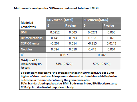
any biologic DMARD(s)]. In multivariable analyses, body
mass index, use of antihypertensives, and rheumatoid nodules, were positively,
while anti-CCP was inversely, correlated with meanSUVtotal and meanSUVmds measurements.
For meanSUVtotal and meanSUVmds, 53% and 59% of the explainable variability in
the measures, respectively, were accounted for by RA factors. In separate
analyses, higher aortic FDG uptake was significantly
correlated with higher echocardiographic E/E’ (a measure of diastolic
dysfunction), and lower end-diastolic and stroke volumes (data not shown).
Conclusion: FDG aortic wall uptake can be performed and
reproducibly measured in relatively large numbers of RA patients, and may serve
as a feasible surrogate measure for CVD in future RA clinical trials. These analyses
suggest that both conventional CV risk factors and RA disease characteristics
are independently associated with FDG uptake in the aorta.
To cite this abstract in AMA style:
Bag Ozbek A, Giles J, Weinberg R, Kinkhabwala M, Zartoshti A, Bokhari S, Bathon J. Assessment of 18F-Fluoro-Deoxyglucose Uptake in the Ascending Aorta of Rheumatoid Arthritis Patients [abstract]. Arthritis Rheumatol. 2015; 67 (suppl 10). https://acrabstracts.org/abstract/assessment-of-18f-fluoro-deoxyglucose-uptake-in-the-ascending-aorta-of-rheumatoid-arthritis-patients/. Accessed .« Back to 2015 ACR/ARHP Annual Meeting
ACR Meeting Abstracts - https://acrabstracts.org/abstract/assessment-of-18f-fluoro-deoxyglucose-uptake-in-the-ascending-aorta-of-rheumatoid-arthritis-patients/

