ACR Meeting Abstracts

ACR Meeting Abstracts

  • Meetings
    • ACR Convergence 2024
    • ACR Convergence 2023
    • 2023 ACR/ARP PRSYM
    • ACR Convergence 2022
    • ACR Convergence 2021
    • ACR Convergence 2020
    • 2020 ACR/ARP PRSYM
    • 2019 ACR/ARP Annual Meeting
    • 2018-2009 Meetings
    • Download Abstracts
  • Keyword Index
  • Advanced Search
  • Your Favorites
    • Favorites
    • Login
    • View and print all favorites
    • Clear all your favorites
  • ACR Meetings

Abstract Number: 1430

A Proof-of-concept Study Evaluating the Use of Functional Brain Magnetic Resonance Imaging in Assessing Treatment Response in Psoriatic Arthritis Patients

Esther Espartal López1, Xabier Michelena Vegas2, Sara Marsal Barril1, Alex Rovira3, Deborah Pareto3 and Alba Erra Duran1, 1Vall Hebron University Hospital, Rheumatology Department, Barcelona, Spain, 2Hospital Universitari Vall Hebron, Barcelona, Spain, 3Vall Hebron University Hospital, Section of Neuroradiology (Department of Radiology, IDI), Barcelona, Spain

Meeting: ACR Convergence 2023

Keywords: Anti-TNF Drugs, Brain, Magnetic resonance imaging (MRI), pain, Psoriatic arthritis

  • Tweet
  • Click to email a link to a friend (Opens in new window) Email
  • Click to print (Opens in new window) Print
Session Information

Date: Monday, November 13, 2023

Title: (1412–1441) Spondyloarthritis Including Psoriatic Arthritis – Treatment Poster II: SpA

Session Type: Poster Session B

Session Time: 9:00AM-11:00AM

Background/Purpose: Inflammatory cytokines can alter the way the central nervous system processes pain as shown in patients with rheumatoid arthritis. However, the relationship between TNFα inhibition and pain processing in individuals with psoriatic arthritis (PsA) has not been investigated.
The purpose of this study is to assess the feasibility of functional magnetic resonance imaging (fMRI) paradigm to estimate the respone to pain and the effect of treatment in PsA patients.

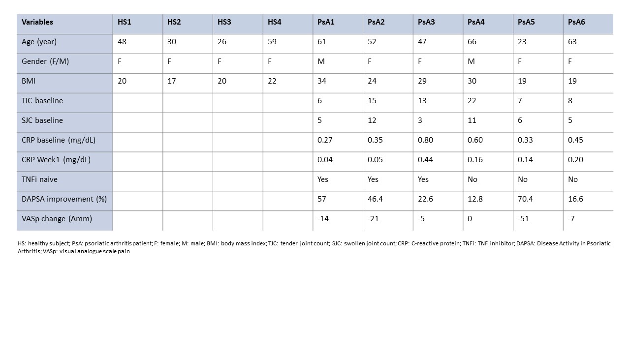
Methods: 6 patients with active PsA eligible to start a TNF inhibitor (TNFi) were included: 3 were naïve to biological disease modifying anti-rheumatic drugs and 3 were experienced. Four healthy subjects (HS) were also included. The physician selected a swollen and tender joint (affected joint) and a control joint in the contralateral hand (non-affected joint). Subjects were scanned in a 3.0T scanner at baseline (BL) and week 1 (W1) after starting treatment. First, the brain pain response was investigated pressing the non-affected joint followed by the affected joint. Each acquisition included 2 fMRI runs. The fMRI effect of pressure was represented for each subject individually, for each time point (p< 0.0001 uncorrected level, minimum extension 10 voxels). We provided a descriptive analysis of the differences in PsA patients brain response between BL and W1 and the clinical response evaluated by the percentage of improvement of Disease Activity in Psoriatic Arthritis (DAPSA) score and visual analogue scale pain (VASp) change.

Results: All subject’s demographic characteristics and PsA patients’ clinical response are shown in Table 1. Brain response to pressure in HS produced activation in the sensory motor cortex, with more or less involvement of additional areas in the prefrontal, parietal and temporal cortex. In PsA patients with a higher treatment response (PsA 1, 2, 5), the brain response to pressure did not show any activation at BL whilst sensory motor cortex activation was seen at W1, with amygdala and insula activation in patients PsA 2 and 5 and prefrontal and frontal activity in patients PsA 1 and 2. In those with less response (PsA 3, 4, 6), the sensory motor cortex was activated at BL with little changes when evaluated at W1 (Fig. 1). No evident differences in brain response activation were observed between naïve and TNFi experienced patients.

Conclusion: The proposed fMRI paradigm seems to be a promising candidate to predict response to treatment with TNFi in PsA patients. Studies with more patients are needed to confirm our preliminary results.

Supporting image 1

Figure 1. Brain fMRI response to pressure of the non-affected joint (green) and the affected joint (red) overlaid on top of the structural image.
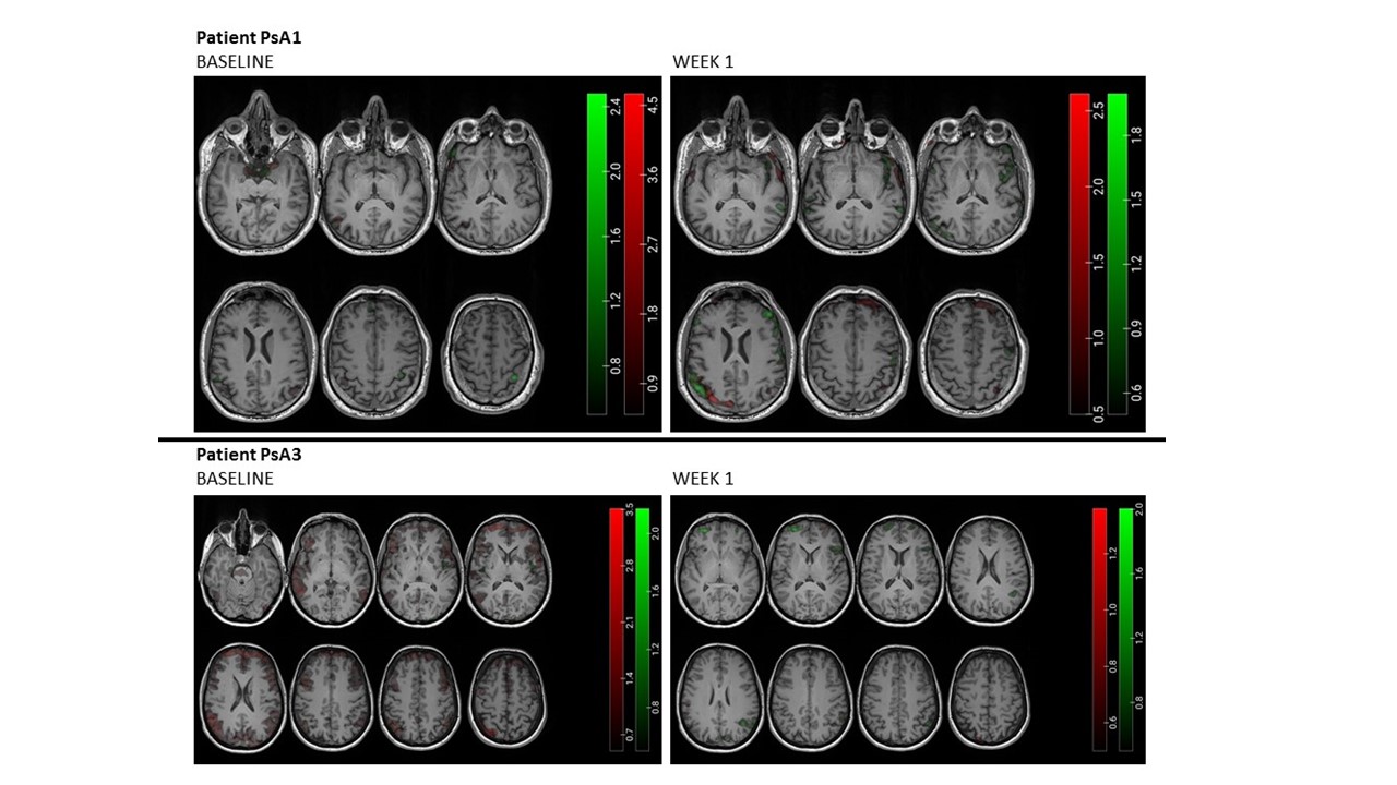
Upper panel corresponds to a patient with clinical improvement (PsA1) and lower panel to a patient that did not respond (PsA3) at baseline (left column) and week 1 (right column).
Differences are observed in the sensorimotor cortex activation between patients: there is an increase in the activation after week 1 in PsA1 while PsA3 showed activation at baseline but not in week 1.

Supporting image 2

Table 1. All subject’s demographic characteristics ans PsA patients’ clinical response


Disclosures: E. Espartal López: Pfizer, 5; X. Michelena Vegas: Pfizer, 5; S. Marsal Barril: Pfizer, 5; A. Rovira: Pfizer, 5; D. Pareto: Pfizer, 5; A. Erra Duran: Pfizer, 5.

To cite this abstract in AMA style:

Espartal López E, Michelena Vegas X, Marsal Barril S, Rovira A, Pareto D, Erra Duran A. A Proof-of-concept Study Evaluating the Use of Functional Brain Magnetic Resonance Imaging in Assessing Treatment Response in Psoriatic Arthritis Patients [abstract]. Arthritis Rheumatol. 2023; 75 (suppl 9). https://acrabstracts.org/abstract/a-proof-of-concept-study-evaluating-the-use-of-functional-brain-magnetic-resonance-imaging-in-assessing-treatment-response-in-psoriatic-arthritis-patients/. Accessed .
  • Tweet
  • Click to email a link to a friend (Opens in new window) Email
  • Click to print (Opens in new window) Print

« Back to ACR Convergence 2023

ACR Meeting Abstracts - https://acrabstracts.org/abstract/a-proof-of-concept-study-evaluating-the-use-of-functional-brain-magnetic-resonance-imaging-in-assessing-treatment-response-in-psoriatic-arthritis-patients/

Advanced Search

Your Favorites

You can save and print a list of your favorite abstracts during your browser session by clicking the “Favorite” button at the bottom of any abstract. View your favorites »

All abstracts accepted to ACR Convergence are under media embargo once the ACR has notified presenters of their abstract’s acceptance. They may be presented at other meetings or published as manuscripts after this time but should not be discussed in non-scholarly venues or outlets. The following embargo policies are strictly enforced by the ACR.

Accepted abstracts are made available to the public online in advance of the meeting and are published in a special online supplement of our scientific journal, Arthritis & Rheumatology. Information contained in those abstracts may not be released until the abstracts appear online. In an exception to the media embargo, academic institutions, private organizations, and companies with products whose value may be influenced by information contained in an abstract may issue a press release to coincide with the availability of an ACR abstract on the ACR website. However, the ACR continues to require that information that goes beyond that contained in the abstract (e.g., discussion of the abstract done as part of editorial news coverage) is under media embargo until 10:00 AM ET on November 14, 2024. Journalists with access to embargoed information cannot release articles or editorial news coverage before this time. Editorial news coverage is considered original articles/videos developed by employed journalists to report facts, commentary, and subject matter expert quotes in a narrative form using a variety of sources (e.g., research, announcements, press releases, events, etc.).

Violation of this policy may result in the abstract being withdrawn from the meeting and other measures deemed appropriate. Authors are responsible for notifying colleagues, institutions, communications firms, and all other stakeholders related to the development or promotion of the abstract about this policy. If you have questions about the ACR abstract embargo policy, please contact ACR abstracts staff at [email protected].

Wiley

  • Online Journal
  • Privacy Policy
  • Permissions Policies
  • Cookie Preferences

© Copyright 2025 American College of Rheumatology