ACR Meeting Abstracts

ACR Meeting Abstracts

  • Meetings
    • ACR Convergence 2024
    • ACR Convergence 2023
    • 2023 ACR/ARP PRSYM
    • ACR Convergence 2022
    • ACR Convergence 2021
    • ACR Convergence 2020
    • 2020 ACR/ARP PRSYM
    • 2019 ACR/ARP Annual Meeting
    • 2018-2009 Meetings
    • Download Abstracts
  • Keyword Index
  • Advanced Search
  • Your Favorites
    • Favorites
    • Login
    • View and print all favorites
    • Clear all your favorites
  • ACR Meetings

Abstract Number: 755

A Novel Diagnostic Algorithm for Polymyalgia Rheumatica Using Three Musculoskeletal Sites on Whole Body PET/CT

Claire Owen1, Aurora Poon 1, Victor Yang 1, Chris McMaster 1, Sze Ting Lee 1, David Liew 1, Jessica Leung 1, Andrew Scott 1 and Russell Buchanan 1, 1Austin Health, Heidelberg, Victoria, Australia

Meeting: 2019 ACR/ARP Annual Meeting

Keywords: 18FDG PET/CT scan, polymyalgia rheumatica and Diagnostic Tests

  • Tweet
  • Click to email a link to a friend (Opens in new window) Email
  • Click to print (Opens in new window) Print
Session Information

Date: Sunday, November 10, 2019

Title: Vasculitis – Non-ANCA-Associated & Related Disorders Poster I: Takayasu's Arteritis & Polymyalgia Rheumatica

Session Type: Poster Session (Sunday)

Session Time: 9:00AM-11:00AM

Background/Purpose: PMR is associated with a pattern of abnormal 18F-Fluorodeoxyglucose (18F-FDG) uptake on whole body PET/CT that is increasingly recognised to correlate anatomically with inflammation of extracapsular structures.(1) The aim of this study was to evaluate the sensitivity and specificity of PET/CT findings in PMR including novel involvement at the posteromedial knee and develop a representative diagnostic scoring algorithm utilising results from a minimum number of musculoskeletal sites.

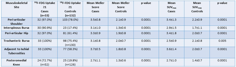
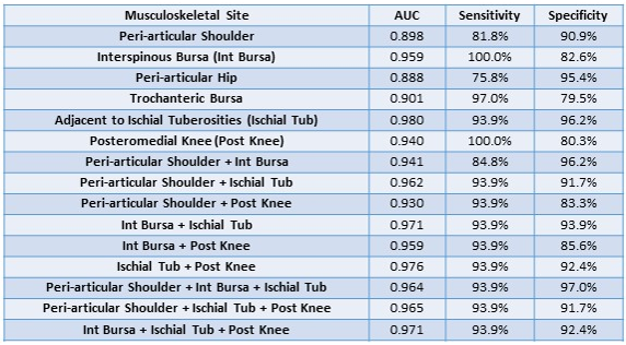
Methods: Steroid-naïve patients with newly diagnosed PMR (2012 EULAR/ACR classification criteria) were prospectively recruited. Participants with giant cell arteritis were excluded. A whole body 18F-FDG PET/CT scan from skull vertex to toes was performed at baseline. Each PMR case was age- and sex-matched to four consecutive historic PET/CT controls, with relevant data pertaining to scan indication, underlying diagnosis and medical history extracted from the clinical record. Qualitative (Meller score) and semi-quantitative scoring (standardised uptake value maximum [SUVmax]) of abnormal 18F-FDG uptake at 21 musculoskeletal sites was undertaken for cases and controls. Results were compared using the Mann-Whitney U test, a p-value < 0.05 being considered statistically significant. Receiver operating characteristics (ROC) curves were generated for abnormal findings at each musculoskeletal site, along with calculation of the area under the curve (AUC), sensitivity and specificity.

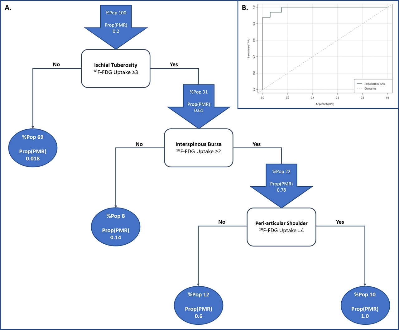
Results: 33 cases met the inclusion criteria and were matched to 132 controls. Mean age was 68.6±7.4 years for cases cf. 68.2±7.3 for controls, and 54.5% of patients were male. Mean EULAR/ACR score was 5.2±0.6 for the PMR group, with median CRP 49 (32 – 65) and ESR 41.5 (24.6 – 64.4). The predominant indication for whole body PET/CT in the controls was malignancy (47.0%). 43/132 controls (32.6%) had a history of a rheumatic condition including 9 (6.8%) with an inflammatory arthritis. Table 1 summarizes the incidence of abnormal findings on whole body PET/CT in cases compared with controls. Individual musculoskeletal sites proved insufficient for diagnostic purposes, however 18F-FDG uptake at the peri-articular shoulder in combination with interspinous bursa and adjacent to the ischial tuberosities achieved a sensitivity of 93.9% and specificity of 97% (Table 2). A diagnostic algorithm utilising scores at each of these 3 musculoskeletal sites is depicted in Figure 1 (AUC 0.987; sensitivity 93.9%, specificity 93.9%). When tested on the study population, none of the controls were misdiagnosed with PMR.              

Conclusion: The combination of abnormal 18F-FDG uptake at the peri-articular shoulder, interspinous bursa and adjacent to the ischial tuberosities on whole body PET/CT is highly sensitive and specific for a diagnosis of PMR.

(1) Owen CE, Poon AMT, Lee ST, Yap LP, Zwar RB, McMenamin CM, et al. Fusion of positron emission tomography/computed tomography with magnetic resonance imaging reveals hamstring peritendonitis in polymyalgia rheumatica. Rheumatology. 2018;57(2):345-53.

Table 1: Incidence, mean Meller score and mean SUVmax for cases compared with controls. Results for continuous variables are reported as mean ± standard deviation.

Table 2: AUC, sensitivity and specificity for PMR diagnosis of abnormal findings at different musculoskeletal sites and their combinations.

Figure 1: A – A diagnostic algorithm for PMR utilising Meller scores at three musculoskeletal sites. %Pop represents the percentage of the total study population with this abnormal finding. Prop-PMR- represents the proportion of PMR cases in that subset; B – ROC curve for the diagnostic algorithm -AUC 0.987; sensitivity 93.9%, specificity 93.9%-.


Disclosure: C. Owen, None; A. Poon, None; V. Yang, None; C. McMaster, None; S. Lee, None; D. Liew, None; J. Leung, None; A. Scott, None; R. Buchanan, None.

To cite this abstract in AMA style:

Owen C, Poon A, Yang V, McMaster C, Lee S, Liew D, Leung J, Scott A, Buchanan R. A Novel Diagnostic Algorithm for Polymyalgia Rheumatica Using Three Musculoskeletal Sites on Whole Body PET/CT [abstract]. Arthritis Rheumatol. 2019; 71 (suppl 10). https://acrabstracts.org/abstract/a-novel-diagnostic-algorithm-for-polymyalgia-rheumatica-using-three-musculoskeletal-sites-on-whole-body-pet-ct/. Accessed .
  • Tweet
  • Click to email a link to a friend (Opens in new window) Email
  • Click to print (Opens in new window) Print

« Back to 2019 ACR/ARP Annual Meeting

ACR Meeting Abstracts - https://acrabstracts.org/abstract/a-novel-diagnostic-algorithm-for-polymyalgia-rheumatica-using-three-musculoskeletal-sites-on-whole-body-pet-ct/

Advanced Search

Your Favorites

You can save and print a list of your favorite abstracts during your browser session by clicking the “Favorite” button at the bottom of any abstract. View your favorites »

All abstracts accepted to ACR Convergence are under media embargo once the ACR has notified presenters of their abstract’s acceptance. They may be presented at other meetings or published as manuscripts after this time but should not be discussed in non-scholarly venues or outlets. The following embargo policies are strictly enforced by the ACR.

Accepted abstracts are made available to the public online in advance of the meeting and are published in a special online supplement of our scientific journal, Arthritis & Rheumatology. Information contained in those abstracts may not be released until the abstracts appear online. In an exception to the media embargo, academic institutions, private organizations, and companies with products whose value may be influenced by information contained in an abstract may issue a press release to coincide with the availability of an ACR abstract on the ACR website. However, the ACR continues to require that information that goes beyond that contained in the abstract (e.g., discussion of the abstract done as part of editorial news coverage) is under media embargo until 10:00 AM ET on November 14, 2024. Journalists with access to embargoed information cannot release articles or editorial news coverage before this time. Editorial news coverage is considered original articles/videos developed by employed journalists to report facts, commentary, and subject matter expert quotes in a narrative form using a variety of sources (e.g., research, announcements, press releases, events, etc.).

Violation of this policy may result in the abstract being withdrawn from the meeting and other measures deemed appropriate. Authors are responsible for notifying colleagues, institutions, communications firms, and all other stakeholders related to the development or promotion of the abstract about this policy. If you have questions about the ACR abstract embargo policy, please contact ACR abstracts staff at [email protected].

Wiley

  • Online Journal
  • Privacy Policy
  • Permissions Policies
  • Cookie Preferences

© Copyright 2025 American College of Rheumatology