Session Information
Date: Tuesday, November 12, 2019
Title: 5T115: SLE – Etiology & Pathogenesis I: Signaling Pathways (2810–2815)
Session Type: ACR Abstract Session
Session Time: 4:30PM-6:00PM
Background/Purpose: In systemic lupus erythematosus (SLE), blood platelets have an activated phenotype, and express high levels of CD40L and P-selectin. Our group demonstrated that SLE patients’ platelets increase type I interferon production by plasmacytoid dendritic cells through CD40-CD40L interaction [1]. Recently, we found that T regulatory cells (Tregs) expressed high levels of P-selectin glycoprotein ligand-1 (PSGL-1), suggesting potential Tregs/platelet interactions.
Methods: Flow cytometry was used for analysis of surface and intracellular molecules expressed on blood cells of active (aSLE, SLEDAI ≥ 6, n= 13 ), inactive (iSLE, SLEDAI < 6, n = 20) SLE patients and healthy donors (HD, n = 17). To assess Tregs immunosuppressive functions, we conducted co-culture experiments of Tregs (CD25high CD127low CD4+) and CFSE-labelled T effector (Teff; CD25–CD127highCD4+) purified from HD. Cells were cultured at a 1:1 ratio with anti-CD3/CD28 activation +/- platelet or recombinant selectin for 6 days. At the end, Teff proliferation was evaluated using cytometry.
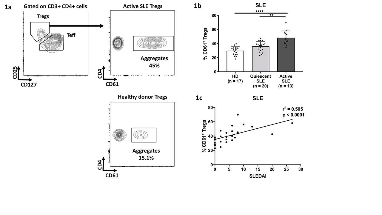
Results: Patients with aSLE had significantly more platelet/Tregs aggregates defined as CD61+ Tregs (figure 1a), than patients with iSLE and HD (figure 1b-c). Conversely, levels of platelet/Teff aggregates were similar in the 3 groups suggesting a specific interaction. To understand the impact of platelet/Tregs interaction, we conducted Teff/Treg co-cultures. Platelets addition to the co-culture inhibited Tregs immunosuppressive functions in a dose-dependent manner. Addition of an anti-PSGL-1 antibody rescued Tregs functions. Lastly, replacing platelets with (P-, E- or L-) selectin could recapitulate these in vitro results.
Mechanistically, P-selectin binding on Tregs induced Syk phosphorylation, which induced a strong intracytosolic calcium release. No similar signals were found with P-selectin binding on Teff. In co-culture assays, Syk inhibition rescued immunosuppressive functions from Tregs exposed to P-selectin.
A transcriptomic analysis of P-selectin-exposed Tregs identified a significant downregulation of the TGF-beta axis. We subsequently confirmed in vitro, by qPCR and cytometry, that P-selectin induced a downregulation of TGF-beta and its chaperone molecule GARP at the RNA and protein levels.
To assess the relevance of selectins in human SLE, we measured levels of soluble selectins (using ELISA) and selectin+ microparticles (by cytometry). Patients with aSLE had increased level of both soluble and microparticular selectins compared to HD and iSLE (p < 0.01 for all comparisons). Microparticular and soluble selectins levels correlated with the SLEDAI (r2 = 0.278, p < 0.001 and r2 = 0.116, p < 0.001, respectively).
Conclusion: The selectin/PSGL-1 axis represents a new pathway responsible for Tregs dysfunction in SLE through downregulation of the TGF-beta axis. Blocking P-selectin (in development for sickle cell disease [2]) might be an innovative treatment option. To evaluate this strategy, we are currently treating DNASE1L3-KO SLE mouse model with an anti-P selectin monoclonal antibody.
References
1 Duffau P, Seneschal J, Nicco C, et al. Sci Transl Med 2010;2:47ra63-47ra63
2 Ataga KI, Kutlar A, Kanter J, et al. N Engl J Med 2017;376:429–39
Fresh whole blood samples from healthy donors or patients with SLE were stained for the study of Tregs/platelet interaction.
-a- Gating strategy to assess Platelet/Tregs circulating aggregates.
-b- Percentage of Platelet/Tregs aggregate in healthy donors or patient with SLE. Quiescent SLE -SLEDAI < 6-, Active SLE -SLEDAI ≥ 6-.**, p < 0.01; ****, p < 0.0001.
-c- Correlation between the percentage of platelet/Tregs aggregates and SLE disease activity as assessed by the SLEDAI.
To cite this abstract in AMA style:
SCHERLINGER M, Guillotin V, Vacher P, Sisirak V, Douchet i, Merillon N, Duffau P, Lazaro E, Ribeiro E, Richez C, BLANCO P. A New Role for Selectins in Systemic Lupus Erythematosus [abstract]. Arthritis Rheumatol. 2019; 71 (suppl 10). https://acrabstracts.org/abstract/a-new-role-for-selectins-in-systemic-lupus-erythematosus/. Accessed .« Back to 2019 ACR/ARP Annual Meeting
ACR Meeting Abstracts - https://acrabstracts.org/abstract/a-new-role-for-selectins-in-systemic-lupus-erythematosus/

