Session Information
Title: Systemic Lupus Erythematosus - Clinical Aspects I - Renal, Malignancy, Cardiovascular Disease
Session Type: Abstract Submissions (ACR)
Background/Purpose:
Patients with lupus are at increased risk for cardiovascular disease (CVD). Abnormalities detected on resting electrocardiography (ECG) that may be associated with an increased risk for subsequent cardiovascular (CV) events include ST-segment abnormalities, T-wave abnormalities, left ventricular hypertrophy (LVH), left-axis deviation, and bundle branch block.
We aimed to describe all abnormalities on resting ECG in a cohort of lupus patients and determine the prevalence of specific abnormalities associated with increased risk for CV events.
Methods:
Resting ECG was performed on all consecutive patients attending The Lupus Clinic between October 2012-May 2013. Participants underwent a standard digitally recorded 12-lead ECG at supine rest. Coded ECGs were reviewed and interpreted by a cardiologist using the Minnesota code classification system.
ECGs were grouped as normal and abnormal. Abnormalities included: pathological Q waves, ST-segment and/or T-wave abnormalities, LVH, left or right bundle branch block, left-axis deviation, arrhythmia and atrial enlargement.
We further determined the prevalence of at least one or more of the ECG findings that may be associated with subsequent CV events: ST-segment and/or T-wave abnormalities, LVH, left-axis deviation, or bundle branch block. We also determined the number of patients who had at least one or more of these variables.
Results:
274 patients were studied. 88.7% of the patients were female with mean age of 47.7 +/- 14.0 and lupus duration of 17.0 +/- 11.5 years.
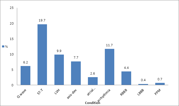
Of 274 resting ECGs 40.5% were abnormal (Figure 1). 21 patients had axis deviation of which 13 had left, 5 had right l and 3 fulfilled criteria for left anterior fascicular block. 7 patients had atrial enlargement (3 right and 4 left). 32 patients with arrhythmia were identified (3 1st degree atrioventricular block, 10 sinus bradycardia, 4 sinus tachycardia, 3 ectopic atrial rhythm, 3 long QTc, 2 isolated premature atrial and 1 isolated premature ventricular contractions, 2 ventricular bigeminy, and 2 short PR interval).
ST-segment abnormalities and/or T-wave abnormalities in 54 (19.7%) LVH in 27 (9.9%), left-axis deviation in 16 (5.8%) and bundle branch block in 13 (4.7%). LBBB were present in 1 patient (0.4%) and in RBBB12 (4.4%). 17 (6.2%) patients had pathological Q waves (which may indicate previous infarct).
115 (41.9%) patients had at least one of the 4 ECG findings that may be associated with subsequent CV events. 23 (8.4%) patients had 2 more ECG abnormalities and 6 (2.2%) were observed to have at least 3 ECG abnormalities.
Conclusion:
40.5% of the ECG had abnormal findings. All of the abnormal ECGs demonstrated at least one ECG finding associated with an increased risk for subsequent CV events. Further studies will determine whether ECG can serve as risk stratification factor for CVD in SLE patients.
Figure 1. Percentage of abnormalities in resting ECG in lupus patients
Disclosure:
Z. Touma,
None;
P. Harvey,
None;
D. D. Gladman,
None;
A. Sabapathy,
None;
M. B. Urowitz,
None.
« Back to 2013 ACR/ARHP Annual Meeting
ACR Meeting Abstracts - https://acrabstracts.org/abstract/lupus-patients-have-a-high-prevalence-of-abnormalities-on-resting-electrocardiogram-that-are-associated-with-increased-risk-for-cardiovascular-events/

