Session Information
Date: Saturday, March 21, 2026
Title: Plenary Abstract Session III
Session Time: 11:15AM-11:30AM
Background/Purpose: Cognitive dysfunction (CD) is a prevalent yet underdiagnosed neuropsychiatric manifestation of childhood-onset systemic lupus erythematosus (cSLE), significantly impacting academic performance, self-management, and overall quality of life. This cross-sectional study employs a multimodal neuroimaging approach to investigate the neurobehavioral basis of CD in cSLE.
Methods: Patients with cSLE and age- and sex-matched healthy controls (HCs) were enrolled in a cross-sectional study to complete questionnaires (Patient-Reported Outcomes Measurement Information System, PROMIS) and multimodal neurocognitive assessments, including the computerized Pediatric Automated Neuropsychological Assessment Metrics (PedANAM) and color-word Stroop task. The Systemic Lupus Erythematosus Disease Activity Index 2000 (SLEDAI-2K) and laboratory markers were used to assess patients’ clinical status. Neuroimaging assessments included functional near-infrared spectroscopy (fNIRS), functional magnetic resonance imaging (fMRI), and structural MRI.
Results: We enrolled Nf20 cSLE patients with a mean age of 17.9 (SD 3.4) and mean disease duration of 51.5 (SD 42.8) months, as well as age and sex-matched HCs (Nf20). Patients with cSLE demonstrated moderate disease activity (mean SLEDAI-2K of 6.5 (SD 5.7). On the PedANAM, cSLE patients vs. HCs demonstrated significantly longer simple reaction times (p = 0.03) as well as poorer performance in the running memory continuous performance test (CPT) (p < 0.01), mathematical processing (p < 0.01), and match-to-sample test (p = 0.02). Relative to HCs, cSLE patients reported worse cognitive functioning (p = 0.02), more psychological stress (p = 0.03) and more physical symptoms (p < 0.01) on PROMIS T-Scores. fNIRS revealed higher prefrontal cortex activity in cSLE vs. HCs during the color-word Stroop task, suggesting impaired cognitive flexibility (Figure 1). fMRI during the N-back working memory task revealed altered frontal cortex and cerebellum activity, while modulations in resting-state fronto-cerebellar connectivity in the cSLE cohort were observed (Figure 2). Moreover, MRIs of patients with cSLE were characterized by reduced gray matter morphological properties in frontal cortex and cerebellar subdivisions (e.g., crus II) alongside altered white matter structural connectivity among these cognitive processing hubs (Figure 3). K-means clustering analysis delineated three subgroups within the cSLE cohort based on neuroimaging profiles, where subgroups were characterized based on PROMIS cognitive functioning (ANOVA Effect Sizes; f = 1.07), depressive symptoms (f = 1.11) and psychological stress (f = 0.69), while standard laboratory and clinical markers of disease severity and activity did not differ between groups.
Conclusion: This study provides evidence of fronto-cerebellar abnormalities and their associations with CD in cSLE. This investigation underscores the need for multidisciplinary research efforts to further elucidate the neurobiological underpinnings of CD in cSLE.
fNIRS Stroop Task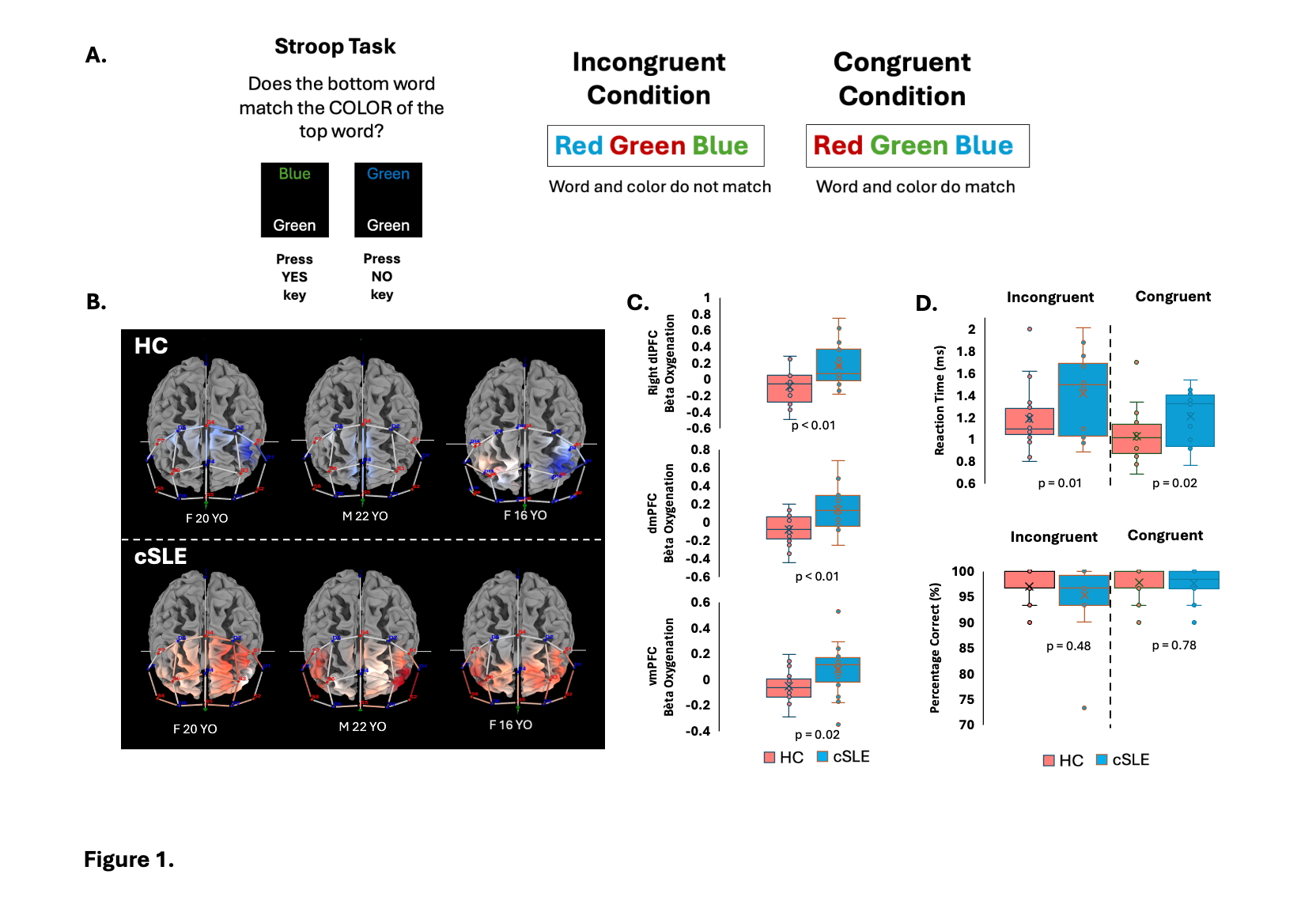 (A) Illustration of the Stroop Task Paradigm, in which participants determined whether the bottom word matches the color of the top word. The task includes congruent (word and color match) and incongruent (word and color do not match) conditions. (B) fNIRS-based contrast (incongruent vs. congruent) activation maps during the Stroop task. Blue represents lower activation, and red represents higher activation, with cSLE participants showing increased prefrontal activation compared to HC. (C) Beta oxygenation values in the right dorsolateral prefrontal cortex (dlPFC), dorsomedial prefrontal cortex (dmPFC), and ventromedial prefrontal cortex (vmPFC), demonstrated significantly higher activation in cSLE participants compared to HC (p = 0.002, p = 0.001, p = 0.015, respectively, two-sided t-tests, corrected for age and sex). (D) Behavioral results showing reaction time (RT) and accuracy for Stroop conditions. cSLE participants exhibited significantly longer RTs in both incongruent (p = 0.01) and congruent (p = 0.02) conditions, while accuracy did not differ significantly between groups.
fMRI Activation and Connectivity.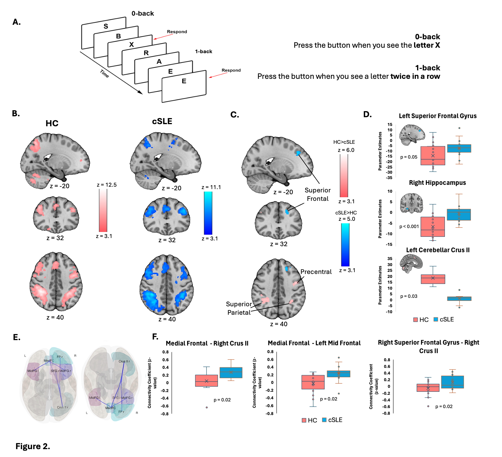 Illustration of the N-back working memory task paradigm. Participants responded when a target letter appeared according to the task condition: in the 0-back condition, participants pressed a button when seeing the letter “X,” while in the 1-back condition, they responded with a button press when a letter repeated consecutively. (B) Within-group average activation maps demonstrating N-Back task activation in HCs (Nf20) and cSLE patients (Nf17) overlayed on the MNI template brain. Mixed-effects analysis, z > 3.1, cluster p-threshold (p=0.05) (C) Significantly greater activation in cSLE patients compared to HCs in superior frontal regions, and decreased activation in the left cerebellar Crus II, right superior parietal cortex, and left precentral gyrus. Mixed-effects analysis, z > 3.1, cluster p-threshold (p=0.05) (D) Subject-specific parameter estimates extracted from 5mm spherical ROIs, showing significantly higher activation in the left superior frontal gyrus, right hippocampus (p = 0.05, p < 0.001 respectively) and lower activation in left cerebellar crus II (p = 0.03,) in cSLE patients compared to HCs. (E) Fronto-Cerebellar Resting-state Functional Connectivity. Frontal and cerebellar Crus II regions overlayed on an inflated template brain surface depicting significantly higher connectivity in patients with cSLE vs. HCs. (p < 0.05, FDR-corrected, corrected for age and sex). Predefined ROIs included bilateral frontal pole, superior and middle frontal gyri, frontal medial cortex and cerebellar Crus II based on the Automated Anatomical Labeling (AAL) Atlas. (F) Bar graph depicting significantly higher connectivity Fisher transformed Z-values for patients with cSLE vs. HCs for (i) medial frontal to right cerebellar Crus II, (ii) medial frontal to left mid frontal and (iii) right superior frontal to right Crus II connectivity. FDR-corrected p-values are shown.
Morphological Alterations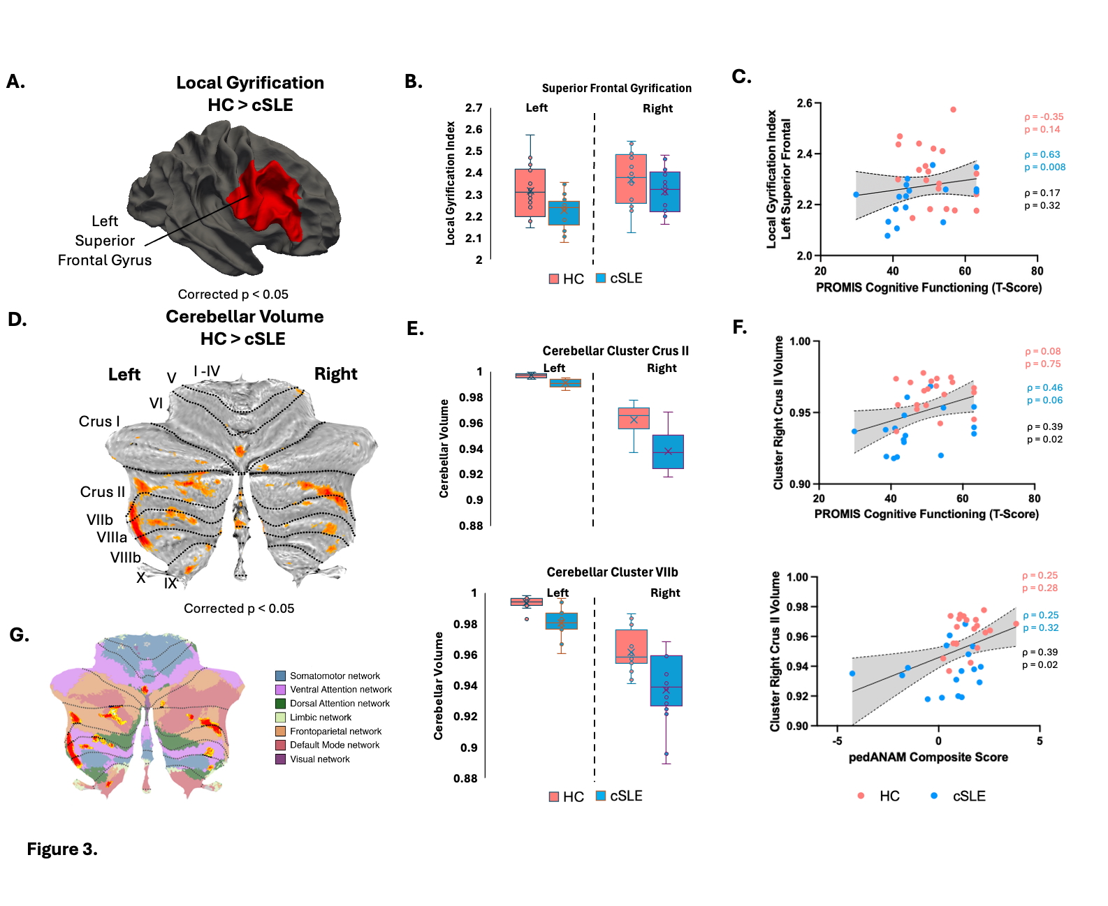 (A) Surface-based analysis revealed significantly lower local gyrification index (LGI) in the left superior frontal gyrus of patients with cSLE vs. HCs (p < 0.05, FDR-corrected). The affected region is visualized on the cortical surface. (B) Box plots depict LGI values in the left and right superior frontal gyrus, showing a significant reduction in patients with cSLE in the left hemisphere but not the right. (C) Scatter plot showing a significant positive correlation between left superior frontal LGI and PROMIS cognitive functioning scores in patients with cSLE (ρ = 0.63, p = 0.008). Spearman Rho (ρ) correlation and p-values for HCs (pink), patients with cSLE patients (blue) and total (black) are shown. (D) Voxel-based morphometry analysis identified significantly reduced cerebellar volume in cSLE patients compared to HCs (FWE-corrected, p < 0.05, cluster-corrected). Affected cerebellar subdivisions include bilateral Crus II, VIIb, VIIIa, among other areas. (E) Box plots displaying reduced cerebellar volume reductions in Crus II and VIIb and in patients with cSLE. (F) Right Crus II volume showed significant positive correlations with PROMIS cognitive functioning (ρ = 0.39, p = 0.02) and pedANAM composite scores (ρ = 0.39, p = 0.02) across all subjects. (G) Overlap of cerebellar volume reductions with functional resting-state gradients, indicating potential functional network implications (adapted from Guell et al., 2018).
To cite this abstract in AMA style:
van der Heijden H, Alonzi G, Cao A, Van Gool R, Koç Yekedüz M, Vrolix L, Ronen I, Rameh V, McBrearty K, Deokar A, Sundel R, Muscal E, Gonzalez-Heydrich J, Knight A, Chang J, Upadhyay J. Fronto-Cerebellar Features Associate with Cognitive Dysfunction in Childhood-Onset Systemic Lupus Erythematosus [abstract]. Arthritis Rheumatol. 2026; 78 (suppl 3). https://acrabstracts.org/abstract/fronto-cerebellar-features-associate-with-cognitive-dysfunction-in-childhood-onset-systemic-lupus-erythematosus/. Accessed .« Back to 2026 Pediatric Rheumatology Symposium
ACR Meeting Abstracts - https://acrabstracts.org/abstract/fronto-cerebellar-features-associate-with-cognitive-dysfunction-in-childhood-onset-systemic-lupus-erythematosus/