Session Information
Session Time: 5:00PM-6:00PM
Background/Purpose: Childhood-onset systemic lupus erythematosus (cSLE) is an inflammatory autoimmune disease that can lead to cognitive dysfunction (CD) across multiple domains. While conventional magnetic resonance imaging can detect brain abnormalities in cSLE, these findings do not always associate with clinical symptoms. Diffusion tensor imaging (DTI) enables detection of subtle white matter (WM) microstructural alterations that may underlie CD, offering potential as a more sensitive approach. This study aimed to: (i) compare global WM DTI metrics between cSLE youth and healthy controls (HCs), and (ii) examine whether global WM DTI metrics differ between cSLE youth with CD, cSLE youth without CD, and HCs.
Methods: Cross-sectional data was acquired from cSLE youth and age-and sex-matched HCs. DTI metrics (fractional anisotropy–FA, mean diffusivity–MD) were computed for global WM. CD was assessed using the Delis-Kaplan Executive Function System (D-KEFS) Color-Word Interference Test based on scaled scores from Color Naming and Word Reading (processing speed), and Inhibition and Inhibition/Switching (executive function) conditions. CD was defined as performance ≥2 standard deviations (SD) below the normative mean in one domain, or ≥1 SD below the mean in two or more domains. A chi-square test examined differences in the proportion of CD between cSLE youth and HCs. Analysis of covariance (ANCOVA), adjusted for age and total WM voxel count, evaluated group differences in FA and MD.
Results: 68 cSLE youth (age 14.9±1.9 years, 85% female) and 62 HCs (age 15.0±1.9 years, 82% female) were included. cSLE youth showed significantly lower FA than HCs (F(1,123)=5.05, p=0.026), with no group differences in MD. CD was present in 17 (25%) cSLE youth and 8 (13%) HCs, with a significantly higher proportion of CD in cSLE youth (χ²(1)=4.27, p=0.039). Among cSLE youth, those with CD had significantly lower FA compared to both non-CD cSLE youth (F(1,62)=4.821, p=0.032) and HCs (F(1,74)=7.923, p=0.006), with no differences in MD. No significant FA or MD differences were found between non-CD cSLE youth and HCs, or between CD and non-CD HCs (all p > 0.05).
Conclusion: cSLE youth demonstrated lower global WM FA compared to HCs. Notably, FA was significantly reduced only in cSLE youth with CD, compared to both non-CD cSLE youth and HCs, suggesting that global WM microstructural alterations may be more pronounced in cSLE youth with cognitive impairment. Future work will examine DTI metrics within specific WM tracts and incorporate additional disease characteristics, such as disease activity and duration.
Table 1. Demographic and Disease Variables.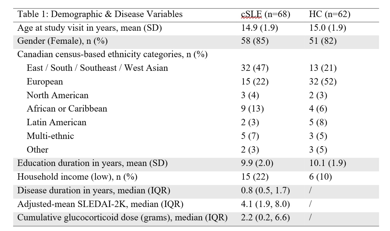
Figure 1. Group differences in global WM DTI metrics among cSLE youth with CD, non-CD cSLE youth, and HCs.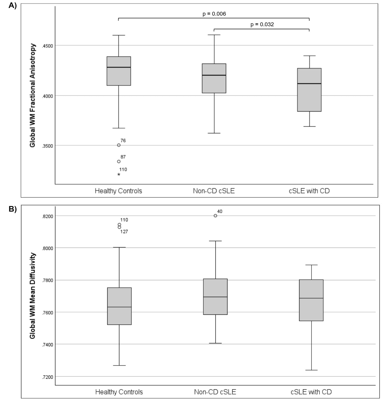 (A) Fractional anisotropy (FA) was lower in cSLE youth with CD compared to both non-CD cSLE youth (F(1,62)=4.821, p=0.032) and HCs (F(1,74)=7.923, p=0.006). (B) Mean diffusivity (MD) showed no significant group differences.
To cite this abstract in AMA style:
Zhou J, Valdes Cabrera D, Mwizerwa O, Jeyanathan A, Ng L, Wagner B, Branson H, Davis A, Levy D, Hiraki L, Knight A. Evaluating Associations between Diffusion Tensor Imaging Metrics and Cognitive Function in Youth with Childhood-Onset Systemic Lupus Erythematosus (cSLE) [abstract]. Arthritis Rheumatol. 2026; 78 (suppl 3). https://acrabstracts.org/abstract/evaluating-associations-between-diffusion-tensor-imaging-metrics-and-cognitive-function-in-youth-with-childhood-onset-systemic-lupus-erythematosus-csle/. Accessed .« Back to 2026 Pediatric Rheumatology Symposium
ACR Meeting Abstracts - https://acrabstracts.org/abstract/evaluating-associations-between-diffusion-tensor-imaging-metrics-and-cognitive-function-in-youth-with-childhood-onset-systemic-lupus-erythematosus-csle/