Session Information
Session Time: 6:00PM-7:00PM
Background/Purpose: Neurologic manifestations in neonatal lupus erythematosus (NLE) are rare and remain poorly characterized. Macrocephaly and hydrocephalus have been reported in approximately 10% of cases, yet these features often go unrecognized or underexplored. While previous studies have described findings from head ultrasonography and brain CT imaging, brain MRI characteristics in NLE have not been reported to date. This gap underscores the need for a deeper understanding of the neuroimaging features associated with the condition.
Methods: We reviewed brain MRI images of two infants diagnosed with NLE and hydrocephalus who were hospitalized at our institution. Both infants demonstrated rapid increases in head circumference (HC), progressing from approximately the 10th percentile at birth to above the 99th percentile at the time of MRI. The mothers of both infants had a confirmed diagnosis of Sjögren’s syndrome. Available head ultrasonography and brain CT images were also analyzed to correlate with the brain MRI findings. Clinical data were reviewed to provide context for the imaging findings and to investigate potential associations with their clinical presentations.
Results: The first brain MRI was performed on a 2-month-old infant who was hospitalized following a rapid progression of HC. The brain MRI revealed diffuse enlargement of the subarachnoid space and all ventricles, with normal brain parenchyma and no abnormal signal in white or grey matter. No evidence of brain atrophy or prior hemorrhage was observed (Figure 1).
The second brain MRI was performed on a 3-month-old infant with history of intrauterine growth restriction (IUGR), born pre-term at 34 weeks gestation, who was hospitalized for respiratory distress and brief cardiac arrest. The brain MRI demonstrated diffuse cerebral white matter atrophy with preserved grey matter, bilateral subdural cerebrospinal (CSF) collections and diffuse enlargement of the subarachnoid spaces. Ventricles were within normal size, and no evidence of acute hemorrhage was observed (Figure2).
Interestingly, the diagnosis of NLE for both infants was confirmed during hospitalization via skin biopsy of their characteristic neonatal lupus rash, which presented at the time of admission. Over time, their HC returned to within the normal growth curve. Both cases are now 5 years old, exhibiting normal developmental milestones with no neurological sequelae.
Conclusion: This study provides the first reported descriptions of brain MRI findings in infants with NLE, highlighting distinct imaging features such as diffuse enlargement of subarachnoid spaces, ventricular abnormalities, and white matter atrophy. These findings expand the understanding of neuroimaging characteristics in NLE. Further research is needed to explore the clinical implications and long-term outcomes of these early neuroimaging abnormalities in NLE.
MRI brain T2 image of a 2-month-old NLE infant with rapid progression of head circumference.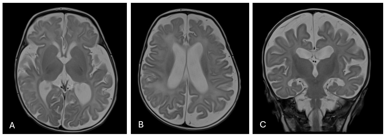 Axial T2 images (A & B) and coronal T2 images (C) demonstrated diffuse enlargement of subarachnoid space and ventricles. Normal signal of grey and white matter.
MRI brain T2 image of a 3-month-old NLE infant, pre-term at 34 weeks gestation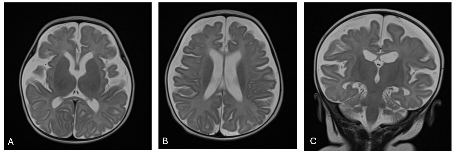 Axial T2 images (A & B) and coronal T2 images (C) demonstrated brain diffuse cerebral white matter atrophy with preserved grey matter, and diffuse enlargement of the subarachnoid spaces. (C) bilateral subdural CSF collections.
To cite this abstract in AMA style:
Panupattanapong S, Zeft A. Novel Insights of Brain MRI Features in Neonatal Lupus Erythematosus with Hydrocephalus [abstract]. Arthritis Rheumatol. 2026; 78 (suppl 3). https://acrabstracts.org/abstract/novel-insights-of-brain-mri-features-in-neonatal-lupus-erythematosus-with-hydrocephalus/. Accessed .« Back to 2026 Pediatric Rheumatology Symposium
ACR Meeting Abstracts - https://acrabstracts.org/abstract/novel-insights-of-brain-mri-features-in-neonatal-lupus-erythematosus-with-hydrocephalus/