Session Information
Date: Thursday, March 19, 2026
Title: Abstracts: Imaging
Session Time: 5:17PM-5:22PM
Background/Purpose: Neuroinflammation is considered to be a major factor in brain-aging—a decline in the structure and function of the brain with age. Presentation of childhood-onset systemic lupus erythematosus (cSLE) coincides with a period of vulnerability in CNS development, highlighting the need for understanding the direct impacts of cSLE on the brain. BrainChart is an interactive, open resource algorithm created to benchmark brain structure and provide a reference dataset indicating age- and sex-based normative neurodevelopmental trajectory across the lifespan. Our study aimed to use BrainChart to examine differences in age- and sex-based neurodevelopmental trajectory as indicated by brain volumes in children with cSLE versus healthy controls (HC).
Methods: We conducted a cross-sectional analysis of baseline structural MRI scans from a prospective cohort of 68 participants with cSLE (meeting ACR, SLICC/ACR or EULAR/ACR classification criteria) aged 11-18 years, and 63 age- and sex-matched HC. Scans were processed using FreeSurfer to calculate grey matter, white matter, subcortical grey matter, and ventricular cerebrospinal fluid (CSF) volumes for each participant. Participant brain volumes, age, sex, and study-specific details were uploaded to BrainChart to benchmark our cohort’s brain volumes against a normative neurodevelopmental trajectory. BrainChart generated centile scores to indicate where each participant’s brain volume falls relative to the normative population. We then used centile scores for each participant to calculate the centile Mahalanobis Distance (CMD) deviation measure, indicating overall neuroanatomical atypicality. We used Mann-Whitney U tests to compare centile scores and CMDs between cSLE and HC groups.
Results: Participant characteristics are shown in Table 1. cSLE participants had a median age of 16.1(IQR 13.8-16.8) years, with a median disease duration of 1.0 (0.6-2.3) years. 2 (3%) of the cSLE participants had a clinical NPSLE diagnosis. HC had a median age of 15.4 (IQR 14.0-17.0). Children with cSLE had significantly lower centile scores for grey matter volume, white matter volume, and subcortical grey matter volume (p < 0.05) compared to HC (Figure 1). Children with cSLE also had significantly higher CMDs compared to HC (p = 0.014) (Figure 2).
Conclusion: Children and adolescents in this cSLE cohort, largely without a clinical NPSLE diagnosis, showed significant deviations from age- and sex-expected norms across multiple brain volumes and higher overall neuroanatomical atypicality. Our results suggest that cSLE may exert widespread neuroanatomical effects affecting the trajectory of neurodevelopment. This emphasizes the need for further longitudinal study of the effects of cSLE on the brain, and attention to brain health in cSLE care.
Table 1: Demographics and clinical characteristics for cSLE and healthy control groups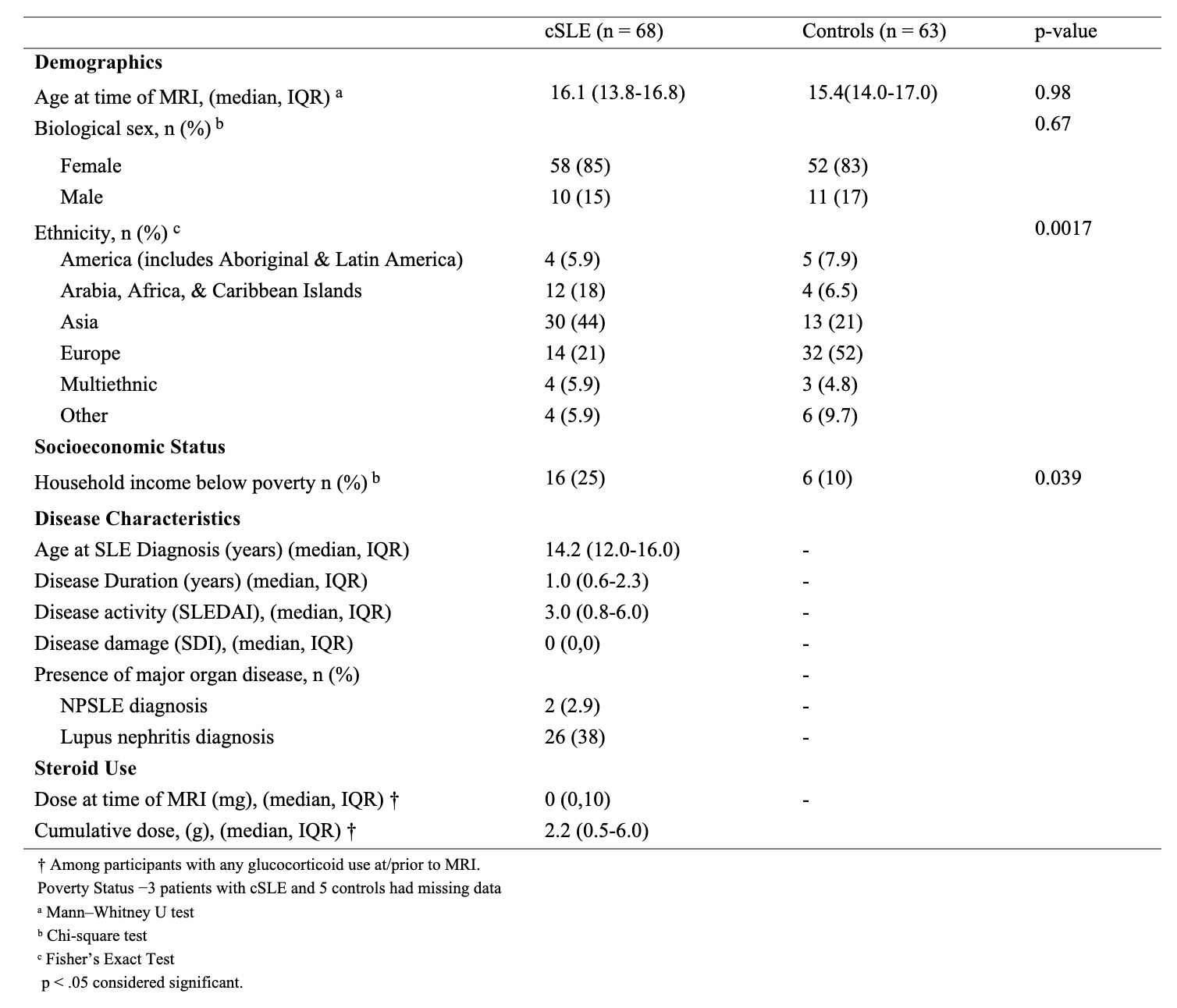 Continuous variables are presented as median (interquartile range), and categorical variables are presented as counts (percentages).
Figure 1: Distribution of global brain volume centile scores by group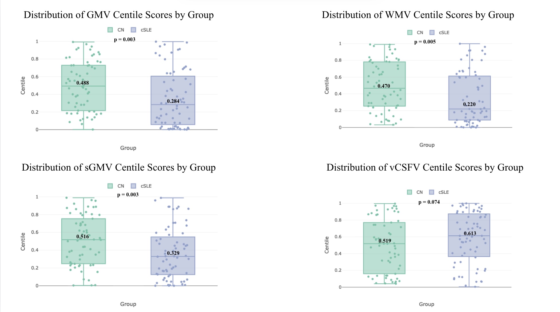 Shown are box plots comparing BrainChart-derived centile scores between children with cSLE and age- and sex- matched HC for grey matter, white matter, subcortical grey matter, and ventricular CSF volumes. Centile scores indicate where one’s brain volume falls relative to the normative population (adjusted for age and sex). A centile score of 0.5 corresponds to the median (expected/typical) value based on age- and sex-based norms. Median centile values for each group are shown within each box, and the p-values listed reflect Mann-Whitney U tests for each comparison.
Figure 2: Distribution of CMDs by group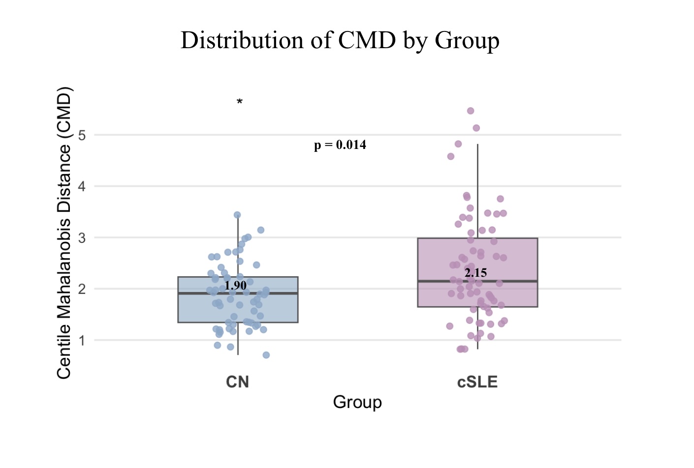 Shown are box plots comparing centile Mahalanobis Distances (CMDs as calculated based on centile scores) between children with cSLE and age- and sex-matched HC. CMD is a multivariate-based composite metric that quantifies the aggregate atypicality of an individual scan; calculated based on the centile scores produced by the 4 global brain volumes. A higher score indicates higher neuroanatomical atypicality.
To cite this abstract in AMA style:
Machado J, Valdes Cabrera D, Branson H, Davis A, Hiraki L, Jeyanathan A, Levy D, Ng L, Wagner B, Knight A. An Examination of Brain-age-related Deviations from Normative Neurodevelopmental Trajectory in Children with Childhood-Onset Systemic Lupus Erythematosus [abstract]. Arthritis Rheumatol. 2026; 78 (suppl 3). https://acrabstracts.org/abstract/an-examination-of-brain-age-related-deviations-from-normative-neurodevelopmental-trajectory-in-children-with-childhood-onset-systemic-lupus-erythematosus/. Accessed .« Back to 2026 Pediatric Rheumatology Symposium
ACR Meeting Abstracts - https://acrabstracts.org/abstract/an-examination-of-brain-age-related-deviations-from-normative-neurodevelopmental-trajectory-in-children-with-childhood-onset-systemic-lupus-erythematosus/