Session Information
Date: Thursday, March 19, 2026
Title: Abstracts: Imaging
Session Time: 5:11PM-5:16PM
Background/Purpose:
Craniofacial localized scleroderma (Cf-LS) is a rare autoimmune disorder that affects face and scalp tissues and is often diagnosed during childhood. Children with Cf-LS may have brain involvement, often ipsilateral to craniofacial lesions, which can be evaluated with magnetic resonance imaging (MRI). However, the extent of disease effects on brain structure beyond lesions is still understudied. We aim to use structural MRI to examine brain morphological abnormalities in patients with Cf-LS compared to healthy controls (HC), and to evaluate if these associate with clinical disease metrics and patient quality of life.
Methods:
Clinical and research T1-weighted brain MRI was retrospectively and prospectively acquired from patients with Cf-LS enrolled in the National Registry for Childhood Onset Scleroderma at the Children’s Hospital of Pittsburgh from 2003-2024 and HC at the Hospital for Sick Children in Toronto, respectively. Left/right lobular cortical grey matter (GM) volume and thickness, lobular white matter (WM) volume, and subcortical GM volumes were automatically segmented in Freesurfer (Fig 1). Group differences on structural brain metrics were evaluated with analysis of covariance and corrected by age, gender, total intracranial volume (ICV) and scanner information. Quantitative brain metrics were compared to a radiological scoring rubric based on routine brain sequences including T2-weighted, susceptibility weighted and diffusion weighted MRIs. Associations between structural metrics, cutaneous disease activity and damage measures (Physician Global Assessment and Localized Scleroderma Cutaneous Assessment Tool), and quality of life (Pediatric Quality of Life Inventory) were evaluated with Pearson correlations adjusted for age and ICV.
Results: Fifty-two patients (22 with right, middle or bilateral sided versus 30 left sided craniofacial lesions) and 30 HC, aged 5-21 years, were included in this study, although groups differed by age (Table 1). Significant group differences were observed in patients versus HC for left/right frontal WM volume and cortical GM volume and thickness (all F >3.37, p < 0.04), left/right parietal cortical GM volume and left parietal cortical GM thickness (all F >3.84, p < 0.026). Lower left subcortical GM volume (R=-0.035, p=0.014) and temporal GM volume (R=-0.032, p=0.025) correlated with greater cutaneous disease damage, while lower occipital WM volume (R=0.052, p=0.004) correlated with worse quality of life in terms of pain and activity.
Conclusion: In this cohort of patients with Cf-LS, we identified brain structural abnormalities beyond regions ipsilateral to craniofacial lesions that were related to greater disease damage and worse quality of life. Future work will assess comparisons with better demographically matched healthy subjects to isolate brain features specific to Cf-LS patients from potential age differences. These initial findings underscore the potential role of structural MRI in monitoring Cf-LS progression in relation to patient outcomes.
Figure 1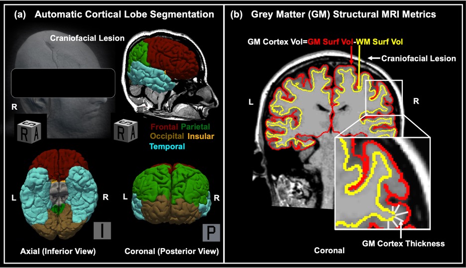 Frontal, parietal, occipital, insular and temporal brain lobes 3D-rendered for the left and right hemispheres in a 17-year-old female patient with a craniofacial lesion in the right side (a). Grey matter (GM) cortical volume was calculated as the volume inside the GM (pial) surface minus the volume inside the white matter (WM) surface in cubic cm and cortical thickness was calculated as the distance (closest point) between the GM and WM surfaces in mm (b). Images were displayed in neurological convention (left hemisphere on the left). Written informed consent to publish has been obtained from this patient.
Table 1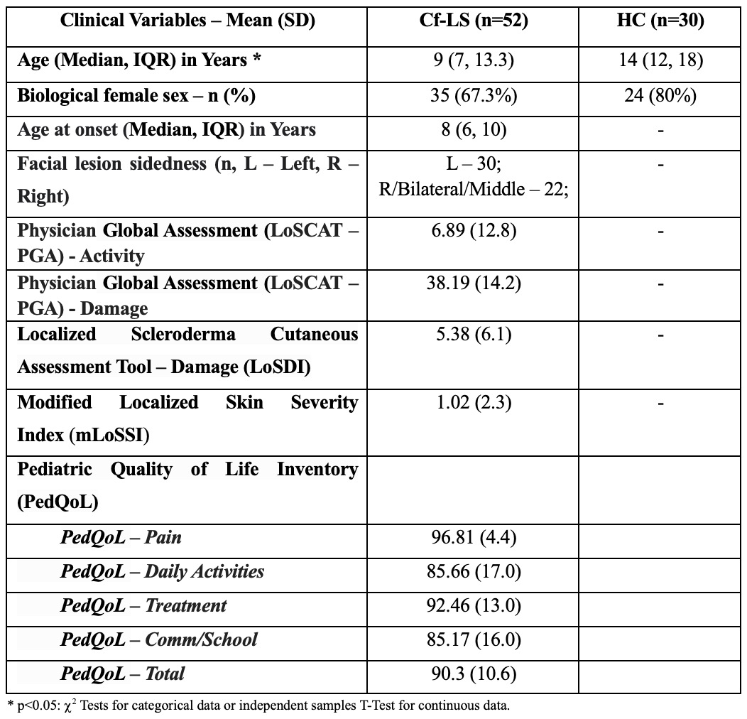 Patient and HC demographics, most frequent Cf-LS manifestations, and clinical scores
To cite this abstract in AMA style:
Valdes Cabrera D, Subramanian S, Glaser D, deRosas E, Havrilla H, Torok K, Knight A. Abnormal Cortical Grey Matter, White Matter and Subcortical Grey Matter Morphometry is Associated with Disease Features and Quality of Life in Patients with Craniofacial Localized Scleroderma [abstract]. Arthritis Rheumatol. 2026; 78 (suppl 3). https://acrabstracts.org/abstract/abnormal-cortical-grey-matter-white-matter-and-subcortical-grey-matter-morphometry-is-associated-with-disease-features-and-quality-of-life-in-patients-with-craniofacial-localized-scleroderma/. Accessed .« Back to 2026 Pediatric Rheumatology Symposium
ACR Meeting Abstracts - https://acrabstracts.org/abstract/abnormal-cortical-grey-matter-white-matter-and-subcortical-grey-matter-morphometry-is-associated-with-disease-features-and-quality-of-life-in-patients-with-craniofacial-localized-scleroderma/