Session Information
Date: Monday, October 27, 2025
Session Type: Poster Session B
Session Time: 10:30AM-12:30PM
Background/Purpose: Axial Spondyloarthritis (axSpA) is a chronic inflammatory disease that mainly involvessacroiliac joints and spine. It is caused by ankylosing spondylitis and other rheumatic diseases and affects mostly young adults, leading to a decreased quality of life and complications if not diagnosed promptly. Conventional radiographs of sacroiliac joints are the first imaging technique in patients with suspected axSpA. However, the diagnosis of sacroiliitis remains subject to significant intrapersonal and interpersonal variation, especially in the early stages. The ability to identify meaningful patterns in large data sets makes machine learning very attractive for screening of diseases such as axSpA.The objective is to To develop and validate a new approach based on artificial intelligence techniques for the automatic classification of the grade of sacroiliitis on conventional radiographs.
Methods: We included 267 X-ray images of patients with clinical suggestion of axSpA. Images were reviewed by 3 rheumatologists and 1 radiologist who graded the severity of sacroiliitis according to the New York criteria (Figure 1 and 2). We performed some pre processing steps using Fiji, median filter, manual adjustment and data augmentation. For model training we used 10-fold stratified cross-validation. Grayscales images were expanded to 3 identical channels and images were resized for the ResNet50, EfficientNetV2S and Xception Backbones. At first we used transfer learning technique by combining a base CNN model as feature extractor, with a top dense stack customized. For the base model, several flavors of Resnet, EfficienNet and Inception architectures were used, besides a custom designed CNN core for basic comparison. We also used XGBoost.
Results: First results were disappointing (Figure 3). To improve performance, we used the XGBoost ensemble learning combined with CNN based onResNet50 architecture. We kept the convolutional kernel, and eliminated the top classifier. A tensor of shape was used as the input for the XGBoost model. After grid search for optimizing XGBoost, we obtained an accuracy of 57% (Figure 3) Therefore, we found that the sensitivity (recall) for all classes was over 60%, except for Class 1 which was misclassified.
Conclusion: -Our trained model successfully detected the grade of sacroiliitis with an accuracy over 60 %.-With further improvement and validation this approach could be helpful to aid the detection and classification of radiographic sacroiliitis
 Examples of pelvic X-rays. A. Normal X-ray. B. Imaging representation of sacroiliitis according to the New York criteria.
.gif) Figure 2. Examples of correct (A) and incorrect (B) results.
Figure 2. Examples of correct (A) and incorrect (B) results.
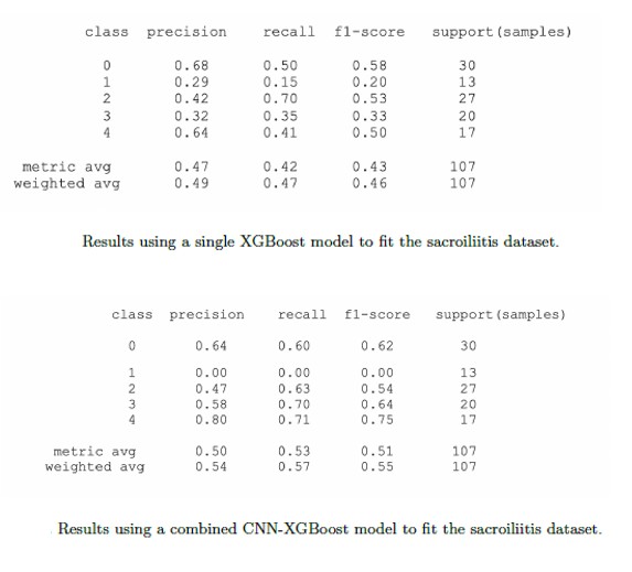 Figure 3. Above initial results using a single XGBoost model to fit the sacroiliitis dataset. Below final results using a combined CNN-XGBoost model to fit the sacroiliitis dataset
To cite this abstract in AMA style:
Fernández Guill E, Bautista Mártir Y, Fernández Jover E, Cases Susarte I. Classification of sacroiliits using an artificial intelligence model [abstract]. Arthritis Rheumatol. 2025; 77 (suppl 9). https://acrabstracts.org/abstract/classification-of-sacroiliits-using-an-artificial-intelligence-model/. Accessed .« Back to ACR Convergence 2025
ACR Meeting Abstracts - https://acrabstracts.org/abstract/classification-of-sacroiliits-using-an-artificial-intelligence-model/