ACR Meeting Abstracts

ACR Meeting Abstracts

  • Meetings
    • ACR Convergence 2024
    • ACR Convergence 2023
    • 2023 ACR/ARP PRSYM
    • ACR Convergence 2022
    • ACR Convergence 2021
    • ACR Convergence 2020
    • 2020 ACR/ARP PRSYM
    • 2019 ACR/ARP Annual Meeting
    • 2018-2009 Meetings
    • Download Abstracts
  • Keyword Index
  • Advanced Search
  • Your Favorites
    • Favorites
    • Login
    • View and print all favorites
    • Clear all your favorites
  • ACR Meetings

Abstract Number: 2650

Extent of Vascular Inflammation on Cranial Vessel Wall MRI and Ophthalmic Complications in Giant Cell Arteritis

David Yang1, Quy Cao2, Fang Liu2, Tara McWilliams2, Ryan Rebello3, Madhura Tamhankar2, Shubhasree Banerjee2, Rui Liang2, Robert Kurtz2, Joshua Baker1, Naomi Amudala2, Zhaoyang Fan4, Jae Song2, Peter Merkel1, Jeffrey Morris2 and Rennie Rhee1, 1University of Pennsylvania, Philadelphia, PA, 2University of Pennsylvania, Philadelphia, 3St Joseph’s Hospital and McMaster University, Hamilton, ON, Canada, 4University of Southern California, Los Angeles, CA

Meeting: ACR Convergence 2024

Keywords: giant cell arteritis, Magnetic resonance imaging (MRI), Vasculitis

  • Tweet
  • Click to email a link to a friend (Opens in new window) Email
  • Click to print (Opens in new window) Print
Session Information

Date: Monday, November 18, 2024

Title: Abstracts: Vasculitis – Non-ANCA-Associated & Related Disorders II

Session Type: Abstract Session

Session Time: 3:00PM-4:30PM

Background/Purpose: There is a need for measures of disease severity for giant cell arteritis (GCA), which may enable identification of high-risk subgroups (e.g., ophthalmic complications) and allow for individualized approach to therapy. Cranial vessel wall MRI (VW-MRI) has potential to quantify disease severity by detecting inflammation of multiple cranial arteries beyond the temporal arteries. We hypothesize a greater extent of vascular inflammation predicts ocular manifestations of GCA, including MRI enhancement of orbital structures, which has been previously demonstrated to potentially detect subclinical orbital disease in GCA. In this study, we derived a continuous score to quantify GCA disease severity based on extent of abnormal enhancement on VW-MRI and assessed the score’s association with: i) diagnosis, ii) ophthalmologic manifestations of GCA and iii) orbital enhancement on MRI.

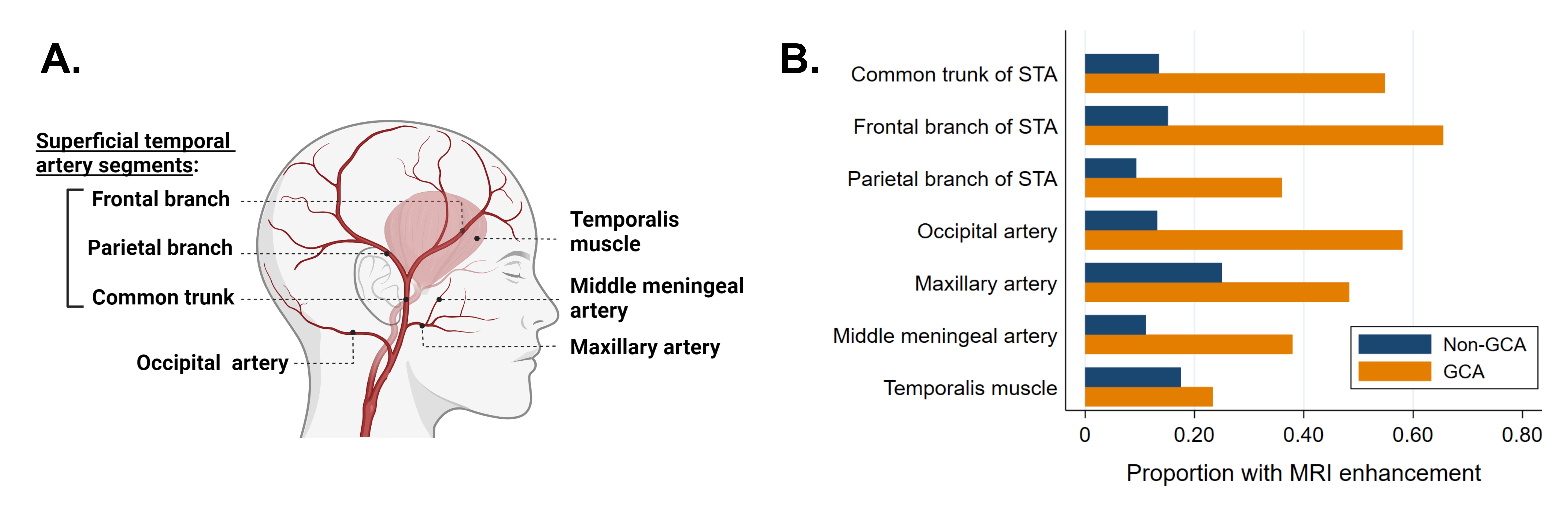
Methods: Patients with suspected new or relapsing GCA who underwent cranial and orbital VW-MRI were included. A subset of patients received repeat MRIs at month 1,6, and 12. Patients were clinically diagnosed as ocular GCA, non-ocular GCA, or non-GCA. A radiologist assessed MRI enhancement of 7 structures bilaterally (Figure 1A). Orbital enhancement on MRI was defined as enhancement of ophthalmic artery or optic nerve sheath. Using a generalized linear mixed effects model with sum-to-zero contrasts, scorable MRI structures (excluding orbital structures) were used to derive a continuous patient-level score representing disease extent. The score, named the Propensity for Enhancement for GCA (PEG) score, ranges from 0 (no enhancement) to 10 (maximum enhancement), accounts for missing arteries, and adjusts for arteries with higher propensities for enhancement. The PEG score was compared between groups using Wilcoxon rank-sum test or linear trend test.


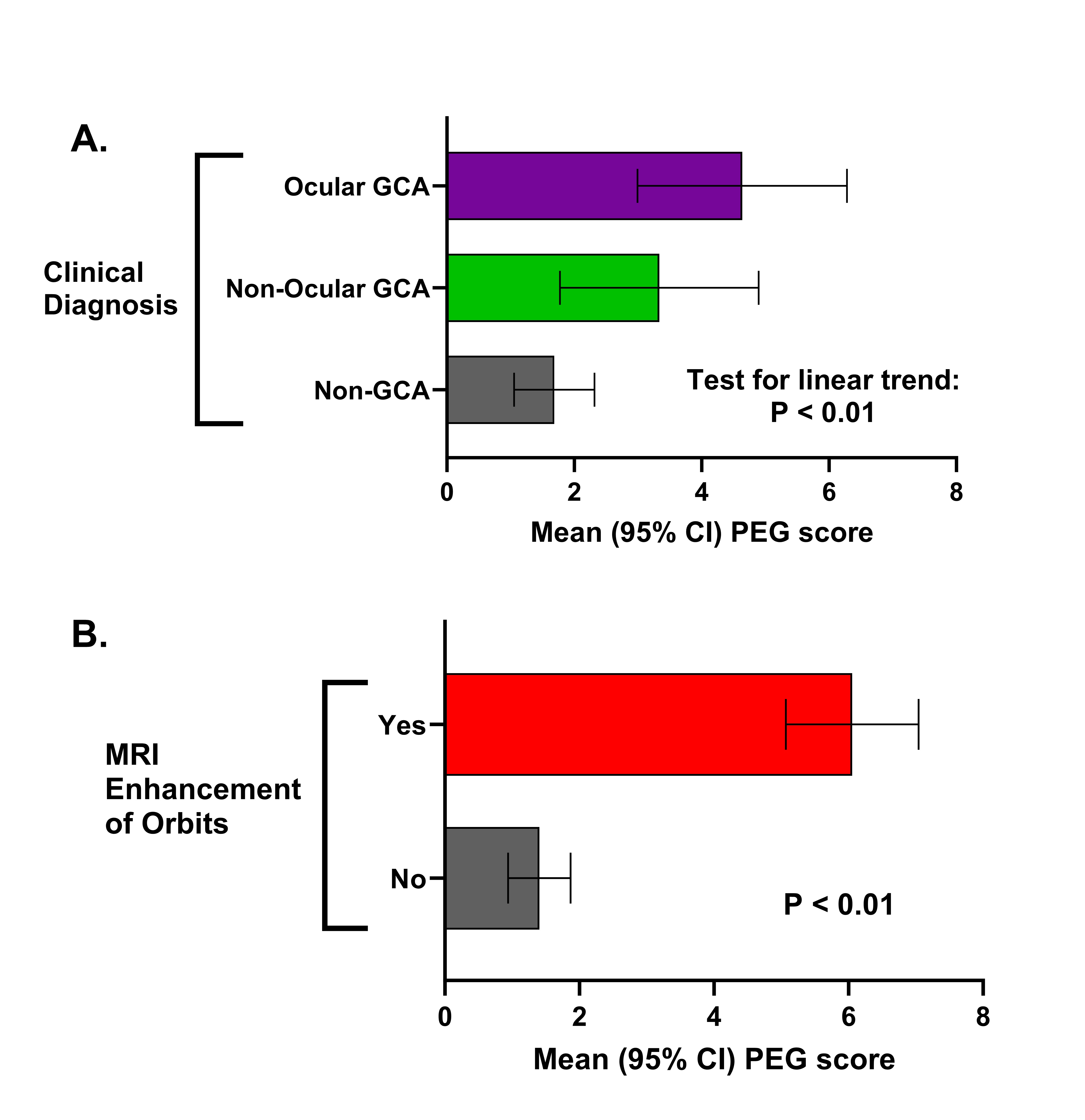
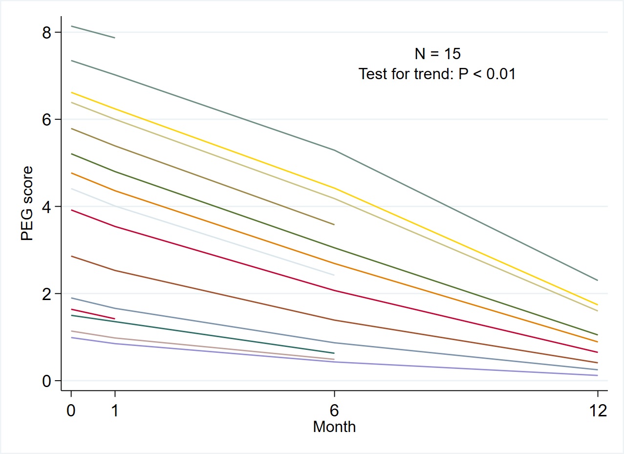
Results: Seventy-five patients (17 ocular GCA, 16 non-ocular GCA, and 42 non-GCA) were included. GCA patients had a greater proportion of structures with abnormal MRI enhancement compared to non-GCA patients. (Figure 1B). PEG score linearly increased across clinical subgroups of non-GCA, non-ocular GCA, and ocular GCA (test for trend: P < 0.01; Figure 2A). Orbital MRI enhancement was observed in 18 (55%) GCA patients including 12 (71%) ocular GCA patients and 6 (38%) with non-ocular GCA. Mean PEG scores were higher in the group with vs. without orbital MRI enhancement (0.61 [0.22] vs. 0.14 [0.17], p< 0.01, Figure 2B). For 15 patients with GCA who underwent repeat MRIs, the PEG score decreased over time with treatment (Figure 3).

Conclusion: VW-MRI reveals the extent of cranial vascular inflammation in GCA patients and is associated with clinical and imaging manifestations of ophthalmic GCA. The PEG score, a novel measure derived from the additive effect of abnormally enhancing MRI structures, is higher in GCA patients with visual manifestations and patients with orbital MRI enhancement. PEG scores improve with treatment. Assessing multiple cranial arteries using VW-MRI may improve assessment of GCA disease severity, which could inform treatment decision-making and clinical trial design.

Supporting image 1

Figure 1. Cranial MRI structures associated with giant cell arteritis. (A) Cranial vasculature and structures associated with giant cell arteritis (GCA) included in models to develop the PEG score. (B) Proportion in each group (GCA vs non-GCA) with abnormal MRI enhancement of each structure.

Supporting image 2

Figure 2. Comparison of global MRI score (PEG score) by ophthalmologic involvement in giant cell arteritis. (A) Mean PEG scores of patients with ocular GCA, non-ocular GCA, and non-GCA (without GCA). (B) Mean PEG scores in patients with versus without abnormal orbital MRI enhancement (defined as optic nerve sheath or ophthalmic artery wall enhancement), which was analyzed due to prior studies hypothesizing that orbital MRI may be an early imaging biomarker of ophthalmic disease in GCA.

Supporting image 3

Figure 3. Change in PEG score with treatment of giant cell arteritis. Lines represent individual PEG scores over time for 15 patients with GCA who received follow-up MRI at 1 month, 6 months, and 12 months after the baseline exam. PEG scores significantly decreased from month 0 to month 6 (P = 0.04) and from month 6 to month 12 (P = 0.03).


Disclosures: D. Yang: None; Q. Cao: None; F. Liu: None; T. McWilliams: None; R. Rebello: None; M. Tamhankar: Amgen, 2, Roche, 2, Viridian, 2; S. Banerjee: None; R. Liang: None; R. Kurtz: None; J. Baker: Cumberland Pharma, 2, Formation Bio, 2, Horizon, 5; N. Amudala: Amgen, 2, 6; Z. Fan: None; J. Song: None; P. Merkel: AbbVie/Abbott, 2, 5, Amgen, 2, 5, argenx, 2, AstraZeneca, 2, 5, Boehringer Ingelheim, 3, 5, Bristol Myers Squibb, 2, 5, Cabaletta, 2, ChemoCentryx, 2, 5, CSL Behring, 2, Dynacure, 2, Eicos, 5, Electra, 5, EMDSerano, 2, Forbius, 2, 5, Genentech/Roche, 2, 5, Genzyme/Sanofi, 2, 5, GSK, 2, 5, HiBio, 2, Immagene, 2, InflaRx, 2, 5, Jannsen, 2, Kiniksa, 2, Kyverna, 2, Magenta, 2, MiroBio, 2, Neutrolis, 2, Novartis, 2, NS Pharma, 2, Pfizer, 2, Q32, 2, 11, Regeneron, 2, Sanofi, 5, Sparrow, 2, 11, Takeda, 2, 5, Talaris, 2, UpToDate, 9, Visterra, 2; J. Morris: None; R. Rhee: None.

To cite this abstract in AMA style:

Yang D, Cao Q, Liu F, McWilliams T, Rebello R, Tamhankar M, Banerjee S, Liang R, Kurtz R, Baker J, Amudala N, Fan Z, Song J, Merkel P, Morris J, Rhee R. Extent of Vascular Inflammation on Cranial Vessel Wall MRI and Ophthalmic Complications in Giant Cell Arteritis [abstract]. Arthritis Rheumatol. 2024; 76 (suppl 9). https://acrabstracts.org/abstract/extent-of-vascular-inflammation-on-cranial-vessel-wall-mri-and-ophthalmic-complications-in-giant-cell-arteritis/. Accessed .
  • Tweet
  • Click to email a link to a friend (Opens in new window) Email
  • Click to print (Opens in new window) Print

« Back to ACR Convergence 2024

ACR Meeting Abstracts - https://acrabstracts.org/abstract/extent-of-vascular-inflammation-on-cranial-vessel-wall-mri-and-ophthalmic-complications-in-giant-cell-arteritis/

Advanced Search

Your Favorites

You can save and print a list of your favorite abstracts during your browser session by clicking the “Favorite” button at the bottom of any abstract. View your favorites »

All abstracts accepted to ACR Convergence are under media embargo once the ACR has notified presenters of their abstract’s acceptance. They may be presented at other meetings or published as manuscripts after this time but should not be discussed in non-scholarly venues or outlets. The following embargo policies are strictly enforced by the ACR.

Accepted abstracts are made available to the public online in advance of the meeting and are published in a special online supplement of our scientific journal, Arthritis & Rheumatology. Information contained in those abstracts may not be released until the abstracts appear online. In an exception to the media embargo, academic institutions, private organizations, and companies with products whose value may be influenced by information contained in an abstract may issue a press release to coincide with the availability of an ACR abstract on the ACR website. However, the ACR continues to require that information that goes beyond that contained in the abstract (e.g., discussion of the abstract done as part of editorial news coverage) is under media embargo until 10:00 AM ET on November 14, 2024. Journalists with access to embargoed information cannot release articles or editorial news coverage before this time. Editorial news coverage is considered original articles/videos developed by employed journalists to report facts, commentary, and subject matter expert quotes in a narrative form using a variety of sources (e.g., research, announcements, press releases, events, etc.).

Violation of this policy may result in the abstract being withdrawn from the meeting and other measures deemed appropriate. Authors are responsible for notifying colleagues, institutions, communications firms, and all other stakeholders related to the development or promotion of the abstract about this policy. If you have questions about the ACR abstract embargo policy, please contact ACR abstracts staff at [email protected].

Wiley

  • Online Journal
  • Privacy Policy
  • Permissions Policies
  • Cookie Preferences

© Copyright 2025 American College of Rheumatology