Session Information
Date: Monday, November 9, 2020
Title: Vasculitis – Non-ANCA-Associated & Related Disorders Poster II
Session Type: Poster Session D
Session Time: 9:00AM-11:00AM
Background/Purpose: Giant cell arteritis (GCA) is the most common form of systemic vasculitis in patients aged 50 years and older.1 Visual symptoms as amaurosis and temporary or permanent loss of visual field secondary to optic nerve ischemia are common manifestations.2 The role of ultrasound in diagnosis of GCA is known.3 Transocular ultrasound of the central retinal artery in GCA patients with visual symptoms has not yet been examined.
Methods: Prospective analysis of flow velocities of the central retinal artery in newly diagnosed GCA patients with visual symptoms and eye-healthy controls. Visual symptoms were defined as amaurosis and temporary or permanent loss of visual field. For each eye, peak systolic values (PS) and end-diastolic values (ED) were recorded.
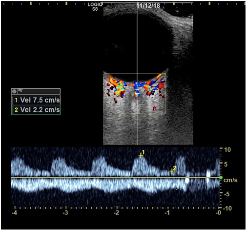
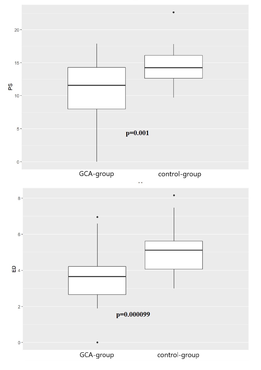
Results: We included 27 newly diagnosed consecutive GCA patients with visual symptoms (GCA-group) and 25 eye-healthy controls. Thirty of 54 eyes (55%) of 27 GCA patients were symptomatic. The control group consisted of 50 central retinal arteries of 25 eye-healthy individuals. Mean age and gender distribution was 75 years (SD± 8.1) with 17 females (63 %) in the GCA group and 67 years (SD± 8.9) with twelve females (48%) in the control group, respectively. Mean flow velocity of the central retinal artery was in 10.9 cm/s (SD± 4.6) in PS and 3.5 cm/s (SD± 1.5) in ED in the GCA group, while values of 14.4 cm/s (SD± 3.2) in PS and 5.1 cm/s (SD± 1.6) in ED were observed in the control group. Mean reduction in flow velocity in the GCA-group was 3.5 cm/s (p-value 0.001) in PS and 1.6 cm/s (p-value 0.000099) in ED, therefore highly significant. Figure 1 shows an ultrasound image of a pathological flow velocity of the central retinal artery in GCA. Figure 2 displays differences in flow velocities between the two groups.
Conclusion: In GCA patients with visual symptoms, a significant reduction of flow velocities of the central retinal artery compared to the eye-healthy control group was observed. Longitudinal data will show, if the flow velocities normalized under treatment.
References
- Warrington KJ, Matteson EL. Management guidelines and outcome measures in giant cell arteritis (GCA). Clin Exp Rheumatol 2007;25:137–41.
- Chean CS, Prior JA, Helliwell T, et al. Characteristics of patients with giant cell arteritis who experience visual symptoms. Rheumatol Int 2019;39:1789–96.
- Dejaco C, Ramiro S, Duftner C, et al. EULAR recommendations for the use of imaging in large vessel vasculitis in clinical practice. Ann Rheum Dis 2018;77:636–43.
 Figure 1. Transocular ultrasound of an affected eye in giant cell arteritis with reduced flow velocities.
 Figure 2: Differences in peak systolic (PS) and end-diastolic (ED) flow between the two groups. GCA-group: Patients with GCA and visual symptoms Control-group: eye-healthy control patients
To cite this abstract in AMA style:
Burg L, Reinking K, Brossart P, Finger R, Behning C, Schaefer V. Prospective Analysis of Flow Velocity of the Central Retinal Artery in Newly Diagnosed Patients with Giant Cell Arteritis with Visual Symptoms and Controls [abstract]. Arthritis Rheumatol. 2020; 72 (suppl 10). https://acrabstracts.org/abstract/prospective-analysis-of-flow-velocity-of-the-central-retinal-artery-in-newly-diagnosed-patients-with-giant-cell-arteritis-with-visual-symptoms-and-controls/. Accessed .« Back to ACR Convergence 2020
ACR Meeting Abstracts - https://acrabstracts.org/abstract/prospective-analysis-of-flow-velocity-of-the-central-retinal-artery-in-newly-diagnosed-patients-with-giant-cell-arteritis-with-visual-symptoms-and-controls/
