Session Information
Date: Tuesday, November 12, 2019
Title: Vasculitis – Non-ANCA-Associated & Related Disorders Poster III: Giant Cell Arteritis
Session Type: Poster Session (Tuesday)
Session Time: 9:00AM-11:00AM
Background/Purpose: PET/CT is a useful modality to diagnose GCA but there is limited knowledge as to how inflammatory vascular findings evolve on follow-up scans. This cohort study aimed to determine the change and clinical significance in vascular wall tracer (FDG) uptake in GCA patients from diagnosis to six-months.
Methods: This study included a subset of patients from the previously reported Giant Cell Arteritis and PET Scan (GAPS) cohort (Sammel et al. Arthritis Rheumatol. 2019). Patients were newly suspected of having GCA and were prospectively enrolled if they had either 1) mural or periadventitial small vessel inflammation on temporal artery biopsy or 2) unequivocal large vessel vasculitis on baseline CT angiogram. Only patients with a clinical diagnosis of GCA were included in the final analysis. PET/CT from the vertex to diaphragm was performed at baseline and at six-months. A single PET-experienced physician, blinded to clinical, biopsy and study inclusion details, reported the grade of vascular FDG uptake for 18 artery segments (0 = none, 1 = minimal/equivocal, 2 = moderate, 3 = very marked). The maximum standard uptake value (SUV) for each vessel was compared to the mean SUV in the left atrium to give an SUVmax/SUVLA ratio. Patients and clinicians were unaware of the six-month PET vascular findings and were followed for 12-months.
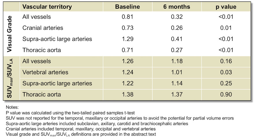
Results: 24/64 GAPS patients met inclusion criteria, of whom 15 consented to six-month PET/CT and had a clinical diagnosis of GCA. From baseline to six-months, the mean vascular uptake grade per patient decreased from 0.81 to 0.32 (Table 1) with significant reductions in all vascular territories (p < = 0.01). The average SUVmax/SUVLA per patient decreased non-significantly from 1.26 to 1.18 (p = 0.16). Over this period the mean CRP fell from 70 to 9 mg/L and ESR fell from 59 to 15 mm/hr. 5/15 (33%) patients had persistent grade 2 (moderate) uptake in at least one artery on the six-month scan. Compared with their counterparts, these 5 patients had a non-significantly higher mean CRP (12 vs 8 mg/L), ESR (16 vs 15 mm/hr) and were taking a lower mean dose of prednisone (8 vs 12 mg). 3 were taking concomitant methotrexate and one an IL-6 inhibitor. None of these 5 patients experienced a clinical flare from 6-12 months but 60% had flares in the preceding six-months.
Conclusion: PET/CT vascular scores decreased in the first six-months following diagnosis of GCA. One-third of patients had significant ongoing vascular uptake at six-months but this did not predict future clinical flare.
To cite this abstract in AMA style:
Sammel A, Hsiao E, Schembri G, Bailey E, Nguyen K, Brewer J, Janssen B, Schrieber L, Youssef P, Fraser C, Laurent R. PET/CT Vascular Findings at Baseline and Six Months in Patients with Newly Diagnosed Giant Cell Arteritis [abstract]. Arthritis Rheumatol. 2019; 71 (suppl 10). https://acrabstracts.org/abstract/pet-ct-vascular-findings-at-baseline-and-six-months-in-patients-with-newly-diagnosed-giant-cell-arteritis/. Accessed .« Back to 2019 ACR/ARP Annual Meeting
ACR Meeting Abstracts - https://acrabstracts.org/abstract/pet-ct-vascular-findings-at-baseline-and-six-months-in-patients-with-newly-diagnosed-giant-cell-arteritis/

