Session Information
Date: Monday, November 11, 2019
Title: Pediatric Rheumatology – ePoster II: SLE, Juvenile Dermatomyositis, & Scleroderma
Session Type: Poster Session (Monday)
Session Time: 9:00AM-11:00AM
Background/Purpose: Kawasaki disease is classically thought to be a monophasic disease that primarily targets the coronary arteries. The American Heart Association Kawasaki guidelines note that patient with severe coronary involvement may have peripheral aneurysms but does not specify which patients to image and how to follow peripheral involvement over time. Hoshino et al followed a cohort of twenty patients with peripheral aneurysms and concluded a diameter greater than 10 millimeters was associated with increased risk of development of stenotic lesions. The risks of additional radiation and sedation to image pediatric patients’ peripheral vessels must be weighed against the benefit of detection peripheral lesions that may cause hemodynamically significant lesions as the patients grow. In this case series, we discuss the presentation and outcome of seven patients found to have peripheral artery aneurysms at a single institution.
Methods: With approval from the Baylor College of Medicine institutional review board, a retrospective review of the medical records of KD patients identified to have peripheral vessel disease was performed. Data including ethnicity, sex, age at diagnosis, total days of fever, day of illness first intravenous immunoglobulin (IVIG) received, coronary aneurysms, peripheral aneurysm characteristics and medications received were abstracted by chart review.
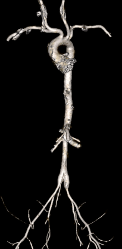
Results: The median age was 9 months, with range of 2 months to 7 years. Most patients described are Hispanic in ethnicity, with one Asian and one African American. Four of the patients described were male and three were female. The median days of fever was 13 days with range of 6 to 34 days. The median day of illness that first IVIG was received was day 10 with range from day 4 to day 34. Six of seven patients had giant coronary aneurysms at some point in the disease course. The peripheral arteries involved were axillary, subclavian, aorta, iliac and femoral. All of the patients received intravenous methylprednisolone in addition to the first IVIG. Three of the seven received a second IVIG, five received infliximab, three received anakinra, and three received cyclophosphamide. Median duration of follow-up was 18 months with range from 2 months to 8 years. Notably, the youngest four of the seven had no coronary aneurysms observed on echo at last follow-up.
Conclusion: Patients described in this cohort had severe disease and often required aggressive multi-modal immunomodulation due to persistent systemic inflammation and extra-coronary involvement. It is reasonable to obtain peripheral imaging in patients that have severe coronary involvement at the second echo or 6-week echo. It is the practice of our center to obtain such imaging when the patients are undergoing cardiac computed tomography. More research is required to understand the frequency of clinically relevant peripheral changes in patients with Kawasaki disease. From this small case series, young patients under 18 months appear to be at increased risk. It is also important to gather information about the normal size of peripheral vessels for very small body sizes. It is possible that this is an under recognized aspect of the disease.
To cite this abstract in AMA style:
Bray M, Ramirez A, Muscal E, De Guzman M. Systemic Vascular Involvement in Kawasaki Disease: A Single Center Cohort [abstract]. Arthritis Rheumatol. 2019; 71 (suppl 10). https://acrabstracts.org/abstract/systemic-vascular-involvement-in-kawasaki-disease-a-single-center-cohort/. Accessed .« Back to 2019 ACR/ARP Annual Meeting
ACR Meeting Abstracts - https://acrabstracts.org/abstract/systemic-vascular-involvement-in-kawasaki-disease-a-single-center-cohort/


