Session Information
Session Type: ACR Abstract Session
Session Time: 2:30PM-4:00PM
Background/Purpose: Gut and joint inflammation in spondyloarthritis (SpA) are closely intertwined. About 50% of axial SpA patients display microscopic signs of inflammation in ileum and/or colon, a risk factor to develop Crohn’s disease over time. It is currently not known if presence of microscopic gut inflammation in new onset SpA is associated with more structural lesions on magnetic resonance imaging of the sacroiliac joints (MRI-SIJ) and whether these lesions relate to the localization of gut inflammation. This study aims to assess whether structural lesions on MRI-SIJ (A) are associated with microscopic gut inflammation in SpA patients and (B) are preferably related to colon or ileum inflammation in case of gut involvement.
Methods: We analyzed baseline information from a patient cohort with a new diagnosis of SpA (expert opinion). Included patients fulfill the Assessment of SpondyloArthritis international Society (ASAS) classification criteria for axial or peripheral SpA. MRI-SIJ was scored by 3 readers, blinded for subject characteristics. Six consecutive slices were assessed for structural lesions: sclerosis, erosions, fatty lesions and (partial) ankylosis. MRI sum scores were analyzed as 2 out of 3 (median) scores. Colon and ileum biopsies were evaluated for microscopic signs of inflammation. The effect of gut inflammation (colon and/or ileum) on MRI-SIJ lesions was investigated by generalized linear models (GLM), adjusted for age and gender and stratified for the SpA phenotype.
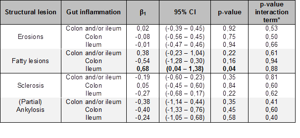
Results: By January 2019, baseline data were available on 105 patients (95 axial and 10 peripheral SpA). Gut inflammation was present in 35 patients (17 ileum, 8 colon, 10 both). Table 1 shows the slope (β1) of the GLMs for erosions, fatty lesions, sclerosis and (partial) ankylosis and the p-value for the SpA phenotype as an interaction term. Erosions, sclerosis, nor ankylosis show a significant association with gut inflammation in general. If present, colon inflammation has no significant relationship with each individual structural lesion. In contrast, presence of ileum inflammation was associated with an increase in the number of fatty lesions by 0,68 (95%CI 0,04 – 1,38). All results are independent of the SpA phenotype (p > 0,05).
Conclusion: Ileal but not colonic inflammatory gut lesions are linked to more fatty lesions on MRI-SIJ in newly diagnosed SpA patients. Baseline microscopic gut inflammation was not associated with erosions, sclerosis nor ankylosis. These data support the concept that microscopic gut inflammation, especially ileal inflammation, is associated with more severe axial inflammation in SpA.
*SpA subtype -axial versus peripheral SpA- as an interaction term.
To cite this abstract in AMA style:
De Craemer A, de Hooge M, Renson T, Deroo L, Lobaton Ortega T, Hoorens A, Carron P, Van den Bosch F, Elewaut D. Ileal but Not Colonic Inflammation Is Linked to Fatty Lesions on MRI of the Sacroiliac Joints in Spondyloarthritis Patients [abstract]. Arthritis Rheumatol. 2019; 71 (suppl 10). https://acrabstracts.org/abstract/ileal-but-not-colonic-inflammation-is-linked-to-fatty-lesions-on-mri-of-the-sacroiliac-joints-in-spondyloarthritis-patients/. Accessed .« Back to 2019 ACR/ARP Annual Meeting
ACR Meeting Abstracts - https://acrabstracts.org/abstract/ileal-but-not-colonic-inflammation-is-linked-to-fatty-lesions-on-mri-of-the-sacroiliac-joints-in-spondyloarthritis-patients/

