Session Information
Session Type: Poster Session (Sunday)
Session Time: 9:00AM-11:00AM
Background/Purpose: FDG-PET-CT is recommended as one of the imaging modalities for the diagnosis and monitoring of primary large-vessel vasculitis (LVV), such as Takayasu’s arteritis (TAK). Interpretation of FDG uptake is based on the principle of determining the inflammatory metabolic activity in the vessel wall in LVV. However, vascular uptake due to atherosclerosis may be a contributing factor in PET-CT assessment. In this study we aimed to evaluate the characteristics of patients with and without involvement of arteries other than the 9 arteries used for assessing PET Vascular Activity Score (PETVAS), which is developed to determine LVV activity in most-commonly involved arterial areas.
Methods: Patients fulfilling ACR1990 classification criteria for TAK and underwent PET-CT imaging were evaluated retrospectively. Demographic and clinical data were collected from patients’ charts. Traditional cardiovascular risk factors: diabetes, hypertension, hyperlipidemia, smoking history and body mass index (BMI) were evaluated. Nine arterial areas used for assessing PETVAS were ascending aorta, aortic arch, descending aorta, abdominal aorta, right and left carotid arteries, innominate artery, right and left subclavian arteries, as originally suggested. Bilateral axillary arteries and iliofemoral arteries were evaluated as atypical (extra-PETVAS) involvement.
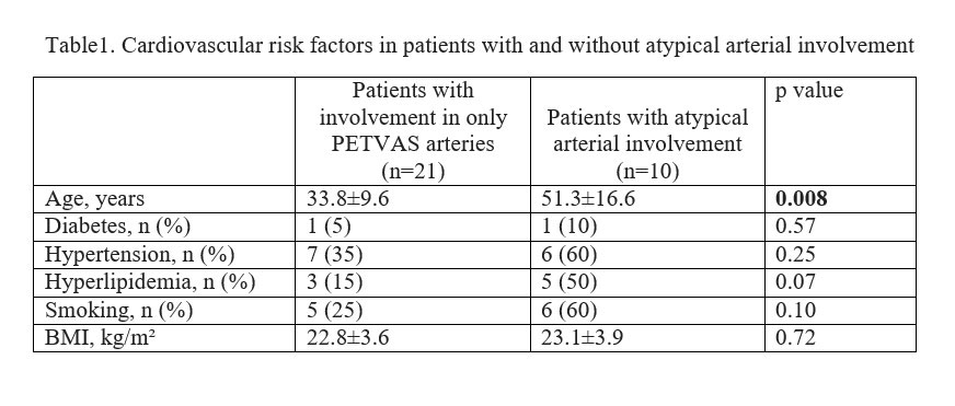
Results: 46 imagings of 34 patients (F/M:28/6) were evaluated. Mean disease duration was 9.3 ±6.5 years and mean age was 40.5± 15.1 years. In the majority of patients aortic arch (87%) was involved followed by ascending aorta (78%) and brachiocephalic artery (50%)(Figure 1). At least one arterial area was involved in 43 images. In 28% of these imagings (12/43), a PET involvement of an artery other than the arteries used for assessing PETVAS, were observed. The mean age of this group was higher than the rest of the group (51.0±16.6 vs 35.8±11.9 years, p=0.01). The number of involved arterial areas (8.5±4.0 vs 3.2±1.5, p=0.000) and total PETVAS scores (10 (2-27) vs 4 (1-17), p=0.013) were higher in atypical arterial involvement group. These patients were also more likely to have the cardiovascular risk factors (Table 1). Patients who had at least 2 cardiovascular risk factors had atypical arterial involvement more frequently (60% (6/10) vs 19% (4/21), p=0.03).
Conclusion: Patients with atypical arterial involvements in PET-CT may have increased atherosclerotic risk factors. These patients had more extended arterial involvement, implying that chronic vascular inflammation may be enhanced by atherosclerosis. Our results suggest that anti-atherosclerotic approaches should be implemented more vigourously in TAK patients.
To cite this abstract in AMA style:
Kaymaz-Tahra S, Ozguven S, Unal A, Alibaz-Oner F, Ones T, Erdil T, Direskeneli H. Involvement of Iliofemoral and Axillary Arteries in PET-CT May Be Associated with Atherosclerotic Risk Factors in Takayasu’s Arteritis [abstract]. Arthritis Rheumatol. 2019; 71 (suppl 10). https://acrabstracts.org/abstract/involvement-of-iliofemoral-and-axillary-arteries-in-pet-ct-may-be-associated-with-atherosclerotic-risk-factors-in-takayasus-arteritis/. Accessed .« Back to 2019 ACR/ARP Annual Meeting
ACR Meeting Abstracts - https://acrabstracts.org/abstract/involvement-of-iliofemoral-and-axillary-arteries-in-pet-ct-may-be-associated-with-atherosclerotic-risk-factors-in-takayasus-arteritis/


