Session Information
Session Type: Poster Session (Sunday)
Session Time: 9:00AM-11:00AM
Background/Purpose: In radiographic axial spondyloarthritis (r-axSpA), whole spine low-dose CT (ldCT) is superior to conventional radiography (CR) in detecting syndesmophytes, mainly due to inclusion of the thoracic spine. Facet joint ankylosis has been studied in r-axSpA in parts of the spine with CR and CT, but not in the whole spine. We aimed to assess readability and interreader reliability of facet joint ankylosis as detected by whole spine ldCT and to describe the prevalence of facet joint ankylosis in each spinal segment in patients with r-axSpA.
Methods: In an observational cohort, r-axSpA patients with syndesmophytes on at least one but no more than 75% of spinal levels of the cervical and lumbar segments on CR and at least one inflammatory lesion on spinal MRI, underwent ldCT (about 4 mSv) of the whole spine. Images were assessed independently by two trained readers and left and right C2-C3 to L5-S1 facet joints were scored as ankylosis present (1) or absent (0). The percentage of missing joint scores due to inability to assess, were calculated per reader and joint level. Joint levels with >15% missing joint scores for ≥1 reader were deemed unreliable to score and were excluded from the study. Interreader reliability was assessed by calculating intraclass correlation coefficients (ICCs), two-way average, absolute agreement. Ankylosis scores were summed per patient per segment and for the whole spine and presented as mean scores per reader. The percentage of patients with an ankylosed joint were presented per joint level in a heatmap.
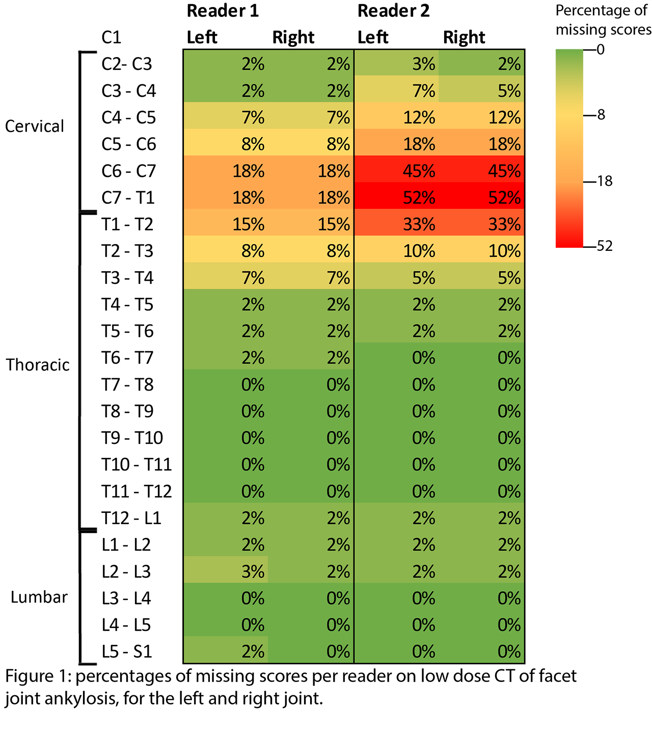
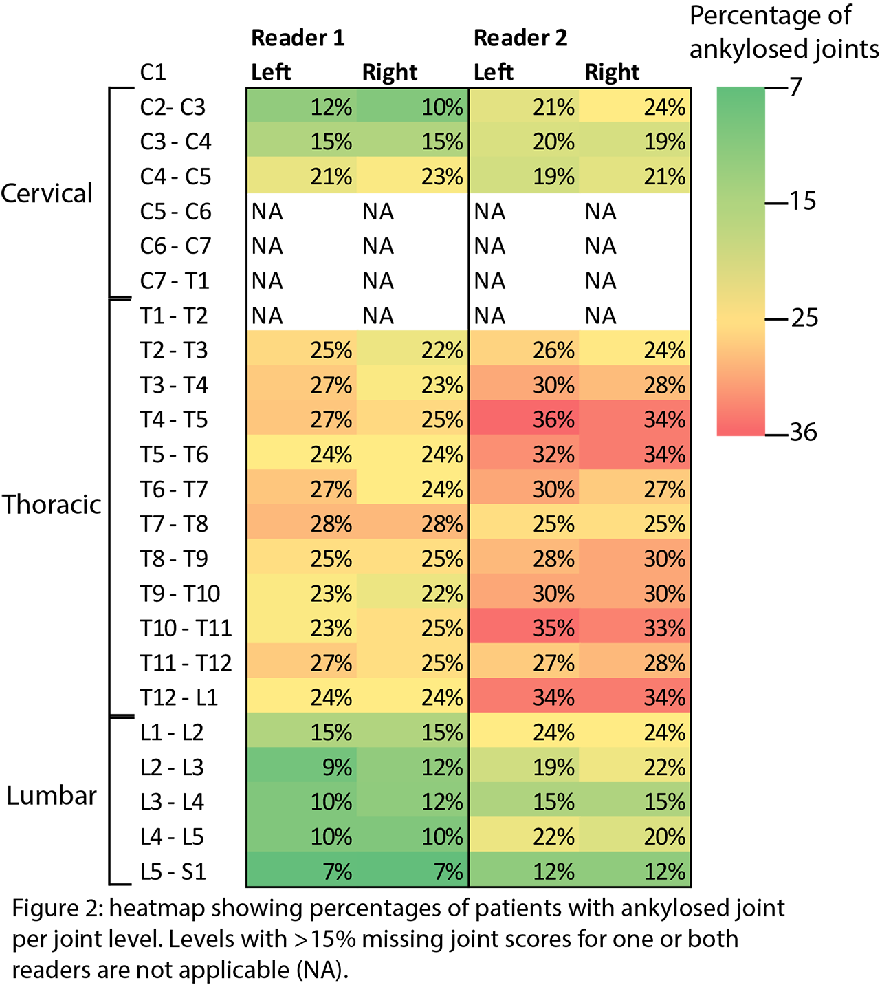
Results: A total of 60 r-axSpA patients were analyzed (mean age 47.7 years, 85% male, 80% HLA-B27+). Reader 1 had between 0% and 18% missing scores per joint, reader 2 had between 0% and 52% missing scores per joint (figure 1). There were >15% missing joint scores for ≥1 reader on levels C5-T2, which were excluded from analyses. Interreader reliability was good to excellent with ICCs ranging from 0.81 to 0.93 (table). Ankylosis occurred at every joint level, but was most prevalent in the thoracic spine (figure 2). Means (SD) of sum-scores for the whole spine and cervical, thoracic and lumbar segments for both readers are presented in the Table.
Conclusion: Facet joints around the cervicothoracic junction were difficult to score in a relatively high percentage of patients and are therefore excluded from scoring. The interreader reliability of the remaining levels was good to excellent. In patients with r-axSpA and at least one syndesmophyte, facet joint ankylosis was detected in all spinal levels, but most ankylosis occurred in the thoracic spine. These results show that ldCT can be used to study facet joint ankylosis in r-axSpA in all spinal segments except the cervicothoracic junction.
To cite this abstract in AMA style:
Stal R, van Gaalen F, Sepriano A, Braun J, Reijnierse M, van der Heijde D, Baraliakos X. Facet Joint Ankylosis on Whole Spine Low-Dose CT in Radiographic Axial Spondyloarthritis [abstract]. Arthritis Rheumatol. 2019; 71 (suppl 10). https://acrabstracts.org/abstract/facet-joint-ankylosis-on-whole-spine-low-dose-ct-in-radiographic-axial-spondyloarthritis/. Accessed .« Back to 2019 ACR/ARP Annual Meeting
ACR Meeting Abstracts - https://acrabstracts.org/abstract/facet-joint-ankylosis-on-whole-spine-low-dose-ct-in-radiographic-axial-spondyloarthritis/


