Session Information
Date: Sunday, November 10, 2019
Title: RA – Diagnosis, Manifestations, & Outcomes Poster I: Risk Factors, Predictors, & Prognosis
Session Type: Poster Session (Sunday)
Session Time: 9:00AM-11:00AM
Background/Purpose: Seropositive arthralgia is defined as joint pain in patients positive for rheumatoid factor (RF) or anti-citrullinated protein antibodies (ACPA). It is a precursor to rheumatoid arthritis (RA) in some individuals. Clinical and synovial findings in seropositive arthralgia when compared to RA and osteoarthritis (OA) have not been fully defined. Our aim was to compare clinical characteristics, macroscopic arthroscopy findings, and synovial immunohistochemical findings in those with seropositive arthralgia, RA, and OA.
Methods: We performed a prospective study of consecutive patients with seropositive arthralgia. All patients were seropositive for RF and/or ACPA. Demographic and clinical characteristics were collected on all patients. Synovial biopsy was performed by needle arthroscopy in all patients, and macroscopic and histologic features recorded. The degree of synovitis and vascularity were recorded on a 0–100-mm visual analog scale. We compared the findings in this group to 2 other cohorts; patients with established diagnoses of either RA or OA as per ACR classification criteria. Whitney U test was used to compare groups. GraphPad Prism Version 8 and IBM SPSS Statistics Version 24 were used for data analysis.
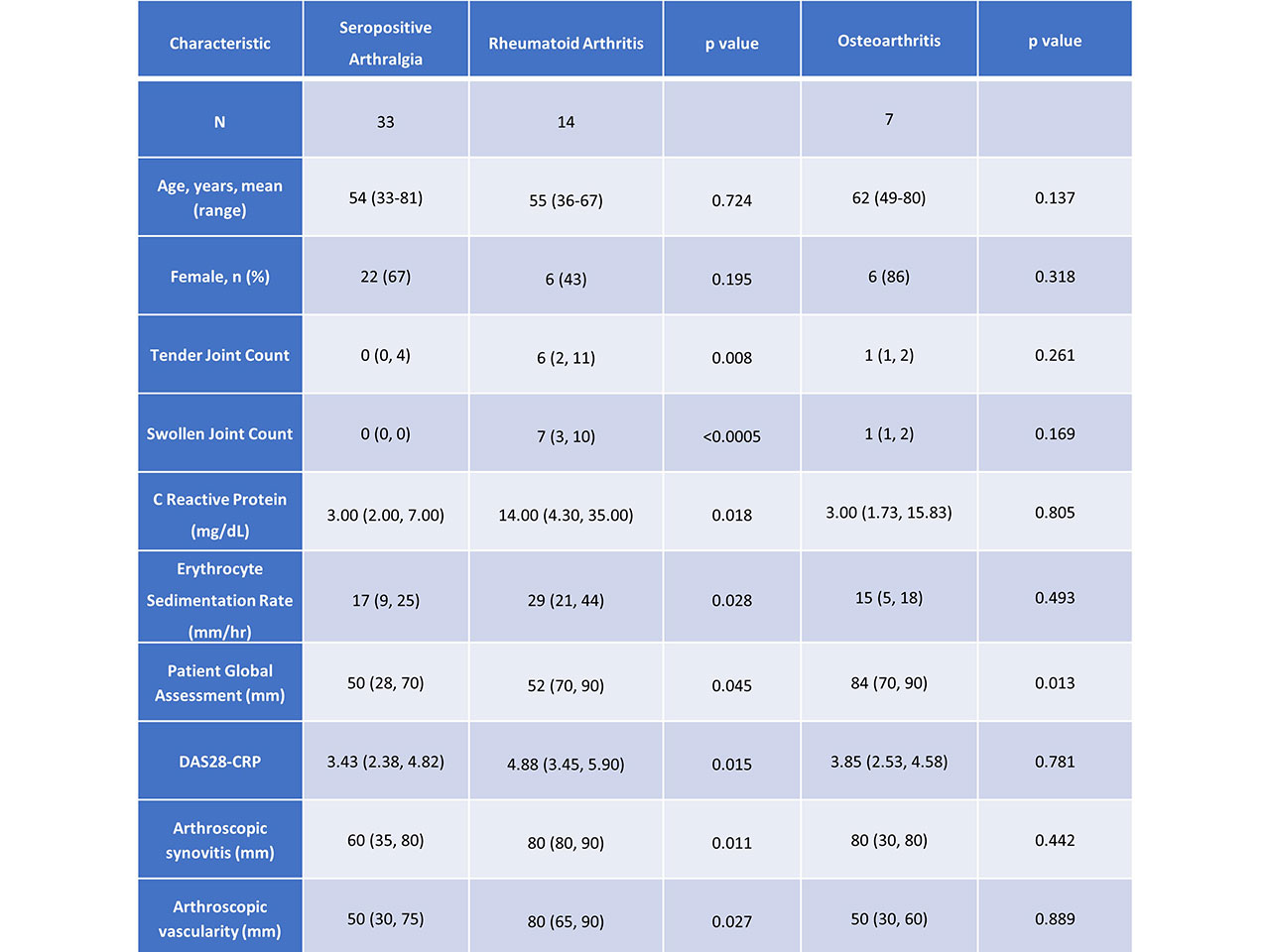
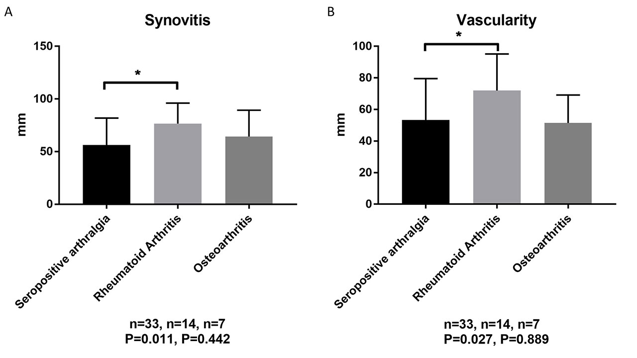
Results: 54 patients were recruited, 33 with seropositive arthralgia, 14 with RA, and 7 with OA. Compared to seropositive arthralgia, established RA patients had higher tender and swollen joint counts, CRP and ESR, patient global assessment (PGA) of disease activity, and DAS28-CRP, Table 1. In contrast, the only difference between seropositive arthralgia and OA patients was higher PGA in the OA group, Table 1. RA patients had significantly greater macroscopic synovitis and synovial vascularity than seropositive arthralgia patients who were not significantly different to OA patients, Table 1, Figure 1. On synovial biopsy, synovial lining layer thickness and microscopic vascularity were increased in RA compared to seropositive arthralgia; there was no difference between seropositive arthralgia and OA. On immunohistochemical analysis of synovial tissue, CD3 and CD38 but not CD138 expression were higher in RA compared to seropositive arthralgia, Figure 2. CD3 and CD38 but not CD138 expression, were higher in seropositive arthralgia compared to OA, Figure 2.
Conclusion: Baseline clinical characteristics, macroscopic arthroscopic findings, and microscopic histologic findings in seropositive arthralgia are more similar to OA than established RA patients. However, key differences in synovial inflammatory cell populations are identifiable in seropositive arthralgia on immunohistochemical analysis.
To cite this abstract in AMA style:
Low C, Conway R, Young F, Molloy E, Mongey A, Wilson G, Fearon U, Veale D. Comparison of Clinical Features, Synovial Histology and Immunohistochemistry in Seropositive Arthralgia, Rheumatoid Arthritis, and Osteoarthritis [abstract]. Arthritis Rheumatol. 2019; 71 (suppl 10). https://acrabstracts.org/abstract/comparison-of-clinical-features-synovial-histology-and-immunohistochemistry-in-seropositive-arthralgia-rheumatoid-arthritis-and-osteoarthritis/. Accessed .« Back to 2019 ACR/ARP Annual Meeting
ACR Meeting Abstracts - https://acrabstracts.org/abstract/comparison-of-clinical-features-synovial-histology-and-immunohistochemistry-in-seropositive-arthralgia-rheumatoid-arthritis-and-osteoarthritis/


