Session Information
Session Type: Abstract Submissions (ACR)
Positron Emission Tomography (PET) using the radiotracer 18F-fluorodeoxyglucose (FDG) with X-ray Computed Tomography (CT) has the ability of producing an in vivo, three-dimensional quantitative map of joint metabolism (hence inflammation) with co-registered anatomy. We report our experience on using 18F-FDG-microPET/CT as a quantitative marker for monitoring the pathogenesis of inflammatory arthritis in the collagen induced arthritis (CIA) mouse model.
Methods:
CIA was induced in 8-12 weeks old male DBA/1 mice (n=10) by injecting 100mg bovine collagen type II in 100ml of complete Freund’s adjuvant intradermally into the proximal tail followed by a second intradermal injection of collagen II with incomplete Freund’s adjuvant on day 21. At baseline (1 day before the first injection), 5 randomly chosen animals underwent microPET/CT scan for assessment of baseline status. At days 28 and 56, 5 animals respectively were scanned using microPET/CT, clinical scoring was performed, and joint tissue was derived for histopathological analysis. Quantitative metrics derived from microPET/CT were correlated against clinical score and histopathology outcomes on a per joint and whole limb basis.
Results:
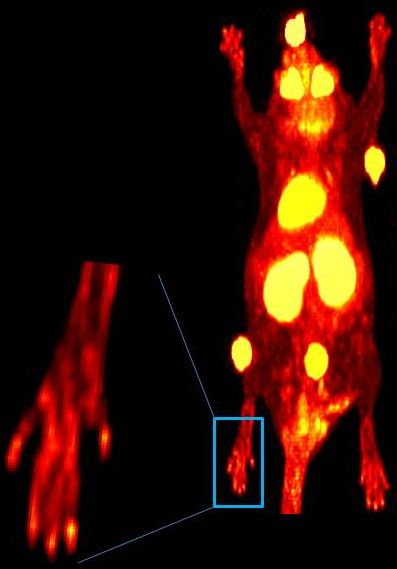
Images from microPET showed a marked increase in metabolic activity at affected joints of the upper and/or lower extremity at follow-up compared to baseline values. microCT images provided detailed assessment of bone destruction. At the early time point (day 28), increase in metabolic activity by an average of ~20% was observed in the limbs. This further increased to an average of ~50% at day 56 as the disease progressed. Focal activity was visualized at individual affected joints by utilizing advanced resolution-recovery methods. microPET/CT metrics at the early and late time points seemed to broadly correlate with clinical and histopathology scores at the arthritic sites.
Conclusion:
Our study shows that quantitative metrics associated with disease pathogenesis can be derived from 18F-FDG-microPET/CT. Changes in clinical score and histopathology outcomes seem to correlate with microPET/CT metrics on both a per joint and whole limb basis. microPET/CT may provide a promising tool for understanding the pathogenesis of arthritis and for monitoring response to new treatments in experimental arthritic models.
Figure 1: An 18F-FDG-microPET scan of the limb of an animal with collagen induced arthritis. Resolution recovery methods allow unparalleled visualization of the details of inflammatory activity.
Disclosure:
S. K. Raychaudhuri,
None;
A. Mitra,
None;
K. Gong,
None;
J. Zhou,
None;
J. Qi,
None;
S. P. Raychaudhuri,
None;
A. J. Chaudhari,
None.
« Back to 2012 ACR/ARHP Annual Meeting
ACR Meeting Abstracts - https://acrabstracts.org/abstract/in-vivo-quantification-of-joint-inflammation-in-a-murine-arthritis-model-by-anato-molecular-imaging/

