Session Information
Session Type: ACR Poster Session B
Session Time: 9:00AM-11:00AM
Background/Purpose: The literature on sonographic enthesitis is primarily based on the large enthesis, ignoring the involvement of the small enthesis such as the hands. In this study, we aimed to determine the prevalence of entheseal abnormalities in small enthesis of the hands in healthy subjects and explore factors that are contributing to the occurrence of these findings.
Methods: Healthy subjects who had no joint pain, recent joint trauma or surgery had US scans of the flexor and extensor tendon insertions to the DIP and extensor tendon insertions to the middle phalanx at the level of the PIP, on the 3rd digits, on both hands. The enthesis were scored as present or absent for elementary lesions of enthesitis (hypoechogenicity, thickening, Doppler signals, enthesophytes, erosions and calcifications) and osteophytes were also recorded.
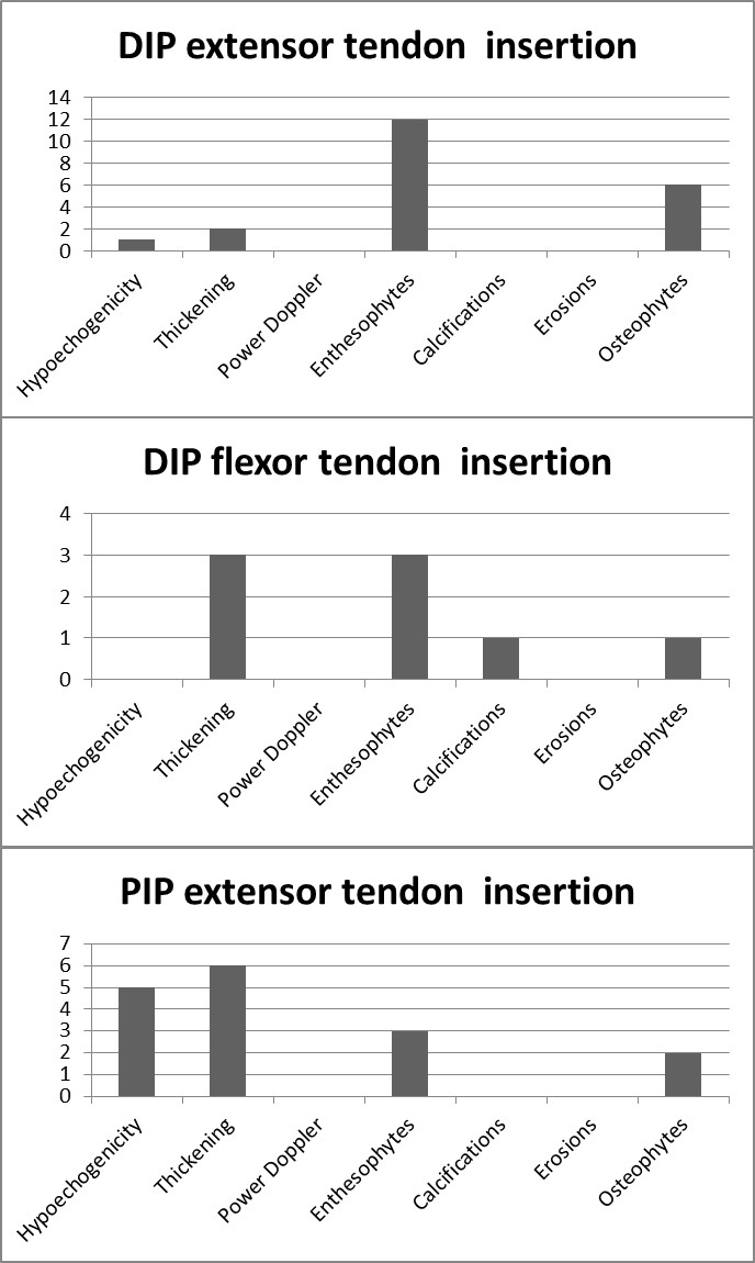
Results: Within 80 healthy subjects (mean age: 45.0 ± 16.1; 62.5% female) 8 had OA. The enthesophytes were the most frequent elementary lesion of enthesitis that could also be seen in the absence of other lesions and were detected in 15% of the DIP extensor tendon insertions, 3.75% of the DIP flexor tendon insertions and 3.75% of the PIP extensor tendon insertions (Figures 1 and 2). 41% of the patients with enthesophytes at the DIP extensor tendon insertion also had osteophytes at the same site, which was significant higher than people without any enthesophytes (5/12 vs 1/68 p<0.001). Patients with enthesophytes were older (67.5 ± 12.6 vs 41.3 ± 13.3; p<0.001) and more frequently men (8/12 vs 22/68; p:0.048). The other elementary lesions were seen in the minority of the digits (Figure 1).
Conclusion: Enthesophytes of the small enthesis are frequent in healthy people and these lesions can be seen in the absence of other elementary lesions. It can be challenging to differentiate enthesophytes from osteophytes at the level of the DIP joints. Therefore the definition of enthesitis for the small enthesis may exclude enthesophytes not to overcall patients with enthesitis. The other features of enthesitis were not common in healthy people, suggesting a good specificity to reflect pathology when detected, however studies on disease groups are needed to clarify.
Figure 1. Elementary lesions of enthesitis on extensor and flexor tendon insertions at the DIP joint and flexor tendon insertion at the PIP joint level.
Figure 2. Longitudinal scans of the extensor tendon insertion to the distal phalanx. A: A healthy enthesis B: Thickening and hypoechogenicity with enthesophyte as well as an osteophyte. MP: Middle Phalanx; DP: Distal Phalanx; o: osteophyte; e: enthesophyte
To cite this abstract in AMA style:
Bakirci S, Solmaz D, Stephenson W, Eder L, Roth J, Aydın SZ. The Evaluation of the Small Enthesis of the Hands By Ultrasound: A Study on Healthy Subjects [abstract]. Arthritis Rheumatol. 2018; 70 (suppl 9). https://acrabstracts.org/abstract/the-evaluation-of-the-small-enthesis-of-the-hands-by-ultrasound-a-study-on-healthy-subjects/. Accessed .« Back to 2018 ACR/ARHP Annual Meeting
ACR Meeting Abstracts - https://acrabstracts.org/abstract/the-evaluation-of-the-small-enthesis-of-the-hands-by-ultrasound-a-study-on-healthy-subjects/


