Session Information
Session Type: ACR Concurrent Abstract Session
Session Time: 9:00AM-10:30AM
Background/Purpose:
Autoantibodies including rheumatoid factor (RF) and antibodies to citrullinated protein antigens (ACPA) are known to be present in the serum of individuals prior to the development of rheumatoid arthritis (RA) (Majka, 2008). These autoantibodies may initially be generated at a mucosal surface given their discovery in the induced sputum of RA and arthritis-free subjects (Willis, 2013). However, there is limited knowledge of the temporal evolution of RF isotypes prior to RA onset, although appearance in the circulation of IgA prior to IgG autoantibody isotypes would support a possible early role for mucosal triggering of autoimmunity in RA.
Methods:
Using the resources of the Department of Defense Serum Repository (DoDSR), we evaluated 214 RA Cases and 158 matched Controls. All RA Case and Control samples were tested for IgA and IgG RF isotypes (Inova). In addition, cut-off for these antibodies were determined using a level that was positive in <5% of a separate group of 58 military controls. The time at which IgA and IgG RF levels differed between RA Cases and Controls prior to diagnosis of RA was evaluated using a linear mixed model after log transformation of IgA and IgG levels. Linear contrasts were used at various times to estimate what time point prior to diagnosis of RA that mean levels of each RF isotype differed between RA Cases and Control, with this analysis serving to identify the earliest time point at which these autoantibodies could be considered abnormal when compared to Controls. For RA Cases, the mean time prior to diagnosis of first positivity of each isotype was also compared using a t-test.
Results:
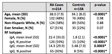
Descriptions of the RA Cases and Controls are presented in the Table. RF-IgA levels were significantly elevated in Cases compared to Controls at 13 years 7 months prior to RA diagnosis. In contrast, RF-IgG levels were significantly higher in Cases compared to Controls at 11 years 1 month prior RA diagnosis. These differences in time of elevation of autoantibodies in Cases compared to Controls, 13 years 7 months vs. 11 years 1 month were statistically significant (p<0.01). Moreover, among RA Cases IgA RF positivity occurred first (mean -1671 days, SD 1761), followed by IgG RF (mean -1359 days, SD 1461) prior to RA diagnosis (p=0.30).
Conclusion:
In analyses of these preclinical RA samples, we found IgA RF to be present prior to the appearance of IgG RF. Given the known relationship of IgA with mucosal inflammation in general, these findings support a potential early mucosal origin for RF generation. In addition, the transition to IgG RF may also play a role in the development of joint symptoms. Future studies should evaluate the role of other factors on the evolution of RF isotypes, as well as the evolution of ACPA isotypes.
To cite this abstract in AMA style:
Kelmenson LB, Wagner BD, Demoruelle MK, Holers VM, Mikuls TR, Edison JD, Deane KD. The Appearance of IgA RF Followed By IgG RF Is Associated with Transition to Classifiable RA in a Large Preclinical RA Cohort [abstract]. Arthritis Rheumatol. 2017; 69 (suppl 10). https://acrabstracts.org/abstract/the-appearance-of-iga-rf-followed-by-igg-rf-is-associated-with-transition-to-classifiable-ra-in-a-large-preclinical-ra-cohort/. Accessed .« Back to 2017 ACR/ARHP Annual Meeting
ACR Meeting Abstracts - https://acrabstracts.org/abstract/the-appearance-of-iga-rf-followed-by-igg-rf-is-associated-with-transition-to-classifiable-ra-in-a-large-preclinical-ra-cohort/

