Session Information
Session Type: ACR Poster Session B
Session Time: 9:00AM-11:00AM
Brain Infiltrating CD4 T Cells Have a Pathogenic T Follicular Helper-Like Phenotype in Neuropsychiatric Lupus
Shweta Jain1, Ariel D Stock2, Fernando Macian-Juan2 and Chaim Putterman1,2
1Division of Rheumatology, Department of Medicine, Albert Einstein College of Medicine and Montefiore Medical Center, Bronx NY 10461.
2 The Departments of Microbiology & Immunology and Pathology, Albert Einstein College of Medicine and Montefiore Medical Center, Bronx NY 10461.
Background/Purpose: The pathogenesis of neuropsychiatric systemic lupus erythematosus (NPSLE) is complex, yet incompletely understood. While the role of T cells is well defined in systemic disease, their involvement in NPSLE has not been carefully studied. Therefore, the aim of this study was to characterize the CD4 T cell populations present in the brain of MRL-lpr/lpr (MRL/lpr) mice, a spontaneous lupus mouse model which exhibits many parallels to human NPSLE.
Methods: T cells infiltrating the brain of female MRL/lpr mice with active neuropsychiatric disease (16-18 weeks of age) were characterized, and subset identification was done by multiparameter flow cytometry, PCR, and cell culture.
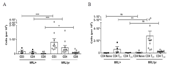
Results: We found extensive infiltration of T cells in the brain of female MRL/lpr mice, almost exclusively limited to the choroid plexus, the site of the brain-CSF barrier. MRL/lpr mice had significant CD4 T cell infiltration as compared to MRL/+ controls (10% of the total cells vs 0.1%, p<0.01). Most of the infiltrating CD4 T cells in MRL/lpr females had an activated phenotype. Specifically, infiltrating CD4 T cells constituted an effector phenotype (80% of CD4+ cells; p<0.01). Importantly, CD4 T cells displayed a T follicular helper cell (TFH) like phenotype, as evidenced by their surface markers (CD4+ICOS+CXCR5+PD1+) and secretion of a signature cytokine, IL-21, in vitro and in vivo. Interestingly, CD4 TFH cells also secreted significant levels of IFN-γ and expressed Bcl-6, thereby conforming to a pathogenic T helper population that drives disease progression. On the other hand, the regulatory axis comprising CD4 T regulatory cells and T follicular regulatory cells was downregulated in MRL/lpr choroid plexus.
 Conclusion: These results imply that accumulation of pathogenic CD4 T follicular helper cells in the brain of MRL/lpr mice may contribute to the neuropsychiatric manifestations of SLE, and suggest this T cell subset as a novel therapeutic candidate.
Figure: T cells infiltrate the choroid plexus of MRL/lpr mice. (A) Single cell suspensions from choroid plexus of 16-18 week old female MRL/+ (n=4) and MRL/lpr (n=10) were stained for the presence of CD3, CD4 and CD8 T cells. (B) CD4 gated cells were characterized as effector (CD4 TEff ; CD4+CD44+), naïve (CD4+CD62L+), and central memory phenotypes (CD4 TCM; CD4+CD44+CD62L+). Values indicate mean±SEM of cells per million and each dot represents one mouse. * p<0.05, ** p<0.01, ***p<0.0001
To cite this abstract in AMA style:
Jain S, Stock A, Macian-Juan F, Putterman C. Brain Infiltrating CD4 T Cells Have a Pathogenic T Follicular Helper-like Phenotype in Neuropsychiatric Lupus [abstract]. Arthritis Rheumatol. 2017; 69 (suppl 10). https://acrabstracts.org/abstract/brain-infiltrating-cd4-t-cells-have-a-pathogenic-t-follicular-helper-like-phenotype-in-neuropsychiatric-lupus/. Accessed .« Back to 2017 ACR/ARHP Annual Meeting
ACR Meeting Abstracts - https://acrabstracts.org/abstract/brain-infiltrating-cd4-t-cells-have-a-pathogenic-t-follicular-helper-like-phenotype-in-neuropsychiatric-lupus/
