ACR Meeting Abstracts

ACR Meeting Abstracts

  • Meetings
    • ACR Convergence 2024
    • ACR Convergence 2023
    • 2023 ACR/ARP PRSYM
    • ACR Convergence 2022
    • ACR Convergence 2021
    • ACR Convergence 2020
    • 2020 ACR/ARP PRSYM
    • 2019 ACR/ARP Annual Meeting
    • 2018-2009 Meetings
    • Download Abstracts
  • Keyword Index
  • Advanced Search
  • Your Favorites
    • Favorites
    • Login
    • View and print all favorites
    • Clear all your favorites
  • ACR Meetings

Abstract Number: 3159

Validation of MRI Structural Lesions Using Computed Tomography in Patients with Axial Spondyloarthritis

Robert G. Lambert1, Damien Loeuille2, Marie Raynal3, Jean Melchior2, Maria Antonietta D'Agostino4, Joel Paschke5 and Walter Maksymowych6, 1Radiology, University of Alberta, Edmonton, AB, Canada, 2Rheumatology, CHRU Vandoeuvre les Nancy, Nancy, France, 3Rheumatology, CHRU Nancy, Nancy, France, 4Rheumatology, Versailles-Saint Quentin en Yvelines University, Boulogne-Billancourt, France, 5CaRE Arthritis, Edmonton, AB, Canada, 6Medicine, University of Alberta, Edmonton, AB, Canada

Meeting: 2016 ACR/ARHP Annual Meeting

Date of first publication: September 28, 2016

Keywords: Computed tomography (CT), magnetic resonance imaging (MRI) and spondylarthritis

  • Tweet
  • Click to email a link to a friend (Opens in new window) Email
  • Click to print (Opens in new window) Print
Session Information

Date: Wednesday, November 16, 2016

Title: Spondylarthropathies and Psoriatic Arthritis – Clinical Aspects and Treatment V: Imaging of Spondyloarthritis

Session Type: ACR Concurrent Abstract Session

Session Time: 9:00AM-10:30AM

Background/Purpose: MRI can detect both inflammatory and structural lesions in the sacroiliac joints (SIJ) of patients with axial SpA. However, standard MRI sequences do not directly depict bone and the appearance of erosion may vary according to the presence/absence of inflammation. Consequently, further validation using an accepted gold standard, namely, computed tomography (CT), is essential. We aimed to assess the detection of structural lesions of bone in the SIJ by MRI using CT as the gold standard.

Methods: CT scans from 44 patients (26 females, mean age 49.4 years, mean symptom duration 9.1 years) were reconstructed in the semicoronal plane parallel to the superior border of the sacrum, as for conventional MRI evaluation of the SIJ, and scoring of lesions by CT was confined to this plane. Structural lesions were scored in consecutive slices in SIJ quadrants (erosion, sclerosis) or SIJ halves (ankylosis) on a dichotomous basis (present/absent) using the same anatomical principles for defining SIJ quadrants as developed for the SPARCC MRI SIJ inflammation and structural scores. Consecutive semicoronal slices from T1W MRI scans of the SIJ from the same cases conducted at the same time as CT were assessed independently for structural lesions (erosion, fat, backfill, ankylosis, sclerosis) blinded to CT assessments using the MORPHO modification of the SPARCC method. MRI lesions were defined according to the Canada-Denmark MRI group. An online scoring module recorded detailed scores for individual SIJ slices. Agreement between CT and MRI was assessed by kappa statistics. Sensitivity/specificity of MRI for CT lesions was calculated. The primary analysis was based on lesions detected concordantly by both readers at the level of the individual iliac/sacral joint surface (erosion, sclerosis) or the individual joint (ankylosis, backfill).

Results: With CT as gold standard and a lesion defined as being present when recorded in the same location on at least 1 coronal slice by both readers, MRI-defined ankylosis had 56.3% sensitivity and 100% specificity for CT ankylosis (Table). For erosion, sensitivity and specificity of MRI was 81.3% and 96.2%, and for sclerosis, sensitivity and specificity of MRI was 50% and 97%, respectively. Agreement between CT and MRI for erosion increased when the cut-off for presence of a lesion was set at 2 slices but only marginally for sclerosis, even when the cut-off for presence of a lesion was set at 3 slices. Lesions observed on CT corresponding to backfill on MRI were ankylosis, erosion, and sclerosis, in 66.7%, 66.7%, and 80% of backfill lesions, respectively.

Conclusion: Ankylosis and erosion on MRI correspond closely with the same type of lesion observed on CT. Both new bone formation and erosion are frequently evident on CT at locations where backfill is observed on MRI supporting the hypothesis that backfill is an intermediary tissue between erosion and ankylosis.  


Disclosure: R. G. Lambert, None; D. Loeuille, None; M. Raynal, None; J. Melchior, None; M. A. D'Agostino, None; J. Paschke, None; W. Maksymowych, Abbvie, 2,Janssen Pharmaceutica Product, L.P., 2,Pfizer Inc, 2,Sanofi-Aventis Pharmaceutical, 2,Abbvie, 5,Boehringer Ingelheim, 5,Eli Lilly and Company, 5,Celgene, 5,Novartis Pharmaceutical Corporation, 5,Pfizer Inc, 5,Sanofi-Aventis Pharmaceutical, 5,UCB, 5,Amgen, 5.

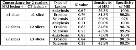
To cite this abstract in AMA style:

Lambert RG, Loeuille D, Raynal M, Melchior J, D'Agostino MA, Paschke J, Maksymowych W. Validation of MRI Structural Lesions Using Computed Tomography in Patients with Axial Spondyloarthritis [abstract]. Arthritis Rheumatol. 2016; 68 (suppl 10). https://acrabstracts.org/abstract/validation-of-mri-structural-lesions-using-computed-tomography-in-patients-with-axial-spondyloarthritis/. Accessed .
  • Tweet
  • Click to email a link to a friend (Opens in new window) Email
  • Click to print (Opens in new window) Print

« Back to 2016 ACR/ARHP Annual Meeting

ACR Meeting Abstracts - https://acrabstracts.org/abstract/validation-of-mri-structural-lesions-using-computed-tomography-in-patients-with-axial-spondyloarthritis/

Advanced Search

Your Favorites

You can save and print a list of your favorite abstracts during your browser session by clicking the “Favorite” button at the bottom of any abstract. View your favorites »

All abstracts accepted to ACR Convergence are under media embargo once the ACR has notified presenters of their abstract’s acceptance. They may be presented at other meetings or published as manuscripts after this time but should not be discussed in non-scholarly venues or outlets. The following embargo policies are strictly enforced by the ACR.

Accepted abstracts are made available to the public online in advance of the meeting and are published in a special online supplement of our scientific journal, Arthritis & Rheumatology. Information contained in those abstracts may not be released until the abstracts appear online. In an exception to the media embargo, academic institutions, private organizations, and companies with products whose value may be influenced by information contained in an abstract may issue a press release to coincide with the availability of an ACR abstract on the ACR website. However, the ACR continues to require that information that goes beyond that contained in the abstract (e.g., discussion of the abstract done as part of editorial news coverage) is under media embargo until 10:00 AM ET on November 14, 2024. Journalists with access to embargoed information cannot release articles or editorial news coverage before this time. Editorial news coverage is considered original articles/videos developed by employed journalists to report facts, commentary, and subject matter expert quotes in a narrative form using a variety of sources (e.g., research, announcements, press releases, events, etc.).

Violation of this policy may result in the abstract being withdrawn from the meeting and other measures deemed appropriate. Authors are responsible for notifying colleagues, institutions, communications firms, and all other stakeholders related to the development or promotion of the abstract about this policy. If you have questions about the ACR abstract embargo policy, please contact ACR abstracts staff at [email protected].

Wiley

  • Online Journal
  • Privacy Policy
  • Permissions Policies
  • Cookie Preferences

© Copyright 2025 American College of Rheumatology