Session Information
Session Type: ACR Poster Session C
Session Time: 9:00AM-11:00AM
Background/Purpose: Assessing disease activity is often challenging in pediatric Takayasu Arteritis (TA). TA inflammation may be clinically silent and not always reflected on laboratory or clinical evaluation. The gold standard for vascular assessment, conventional angiography, cannot always be safely performed. Reliance on other modalities such as contrast-enhanced CT and MRI evaluate anatomy, but may not show the full extent of inflammation. PET/CT offers improved assessment of inflammation but at the expense of radiation exposure and poor anatomic resolution. Our facility is the first stand-alone pediatric hospital in North America with PET/MRI, which offers potential for superior functional and anatomic information with lower radiation exposure relative to stand-alone PET/CT with documentation of a 40% lower dose of radiation of whole body PET/MR relative to PET/CT. We describe cases where PET/MRI influenced pediatric TA patient care.
Methods: With IRB approval we reviewed 18F-flurodeoxyglucose (FDG) PET/MRI on two pediatric patients with TA obtained as part of their clinical care. Anatomic whole body MRI sequences as well as FDG PET were acquired as part of the PET/MRI protocol.
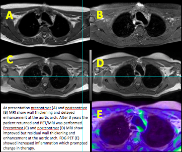
Results: Two adolescents obtained PET/MRI during 2015. A 17-year old female had been in clinical remission (off medications) for two years had newly elevated inflammatory markers. Her contrast MRI was interpreted as showing improved disease activity as compared to prior scans. Corresponding FDG-PET indicated ongoing disease activity in the walls of the aortic arch, right innominate artery, and left common carotid artery. Analysis of lab and PET/MRI data led to re-initiation of infliximab, methotrexate, and corticosteroids. A 12-year old female diagnosed at an outside hospital 3 months earlier and on infliximab, methotrexate, and corticosteroids was transferred in acute heart failure. Her inflammatory markers were normal and clinical exam was unchanged from recent rheumatology evaluation. PET/MRI showed involvement of the right common and internal carotid arteries, the aortic root to the suprarenal abdominal aorta, the right pulmonary artery, as well as hypermetabolism within the pericardial fluid. The disease was more extensive than had recently been shown on conventional imaging and led to initiation of tocilizumab.
Conclusion: PET/MRI may delineate the extent of large vessel inflammation and persistence of disease activity in all stages of pediatric TA evaluation. PET vascular metabolic data may be obtained longitudinally with a reduced radiation burden and may allow for timely targeting of ongoing inflammation.
To cite this abstract in AMA style:
Gillispie M, Rosillo P, De Guzman M, Seghers V, Masand P, Muscal E. Developing a Role for 18f-Flurodeoxyglucose PET/MRI in Pediatric Takayasu Arteritis [abstract]. Arthritis Rheumatol. 2016; 68 (suppl 10). https://acrabstracts.org/abstract/developing-a-role-for-18f-flurodeoxyglucose-petmri-in-pediatric-takayasu-arteritis/. Accessed .« Back to 2016 ACR/ARHP Annual Meeting
ACR Meeting Abstracts - https://acrabstracts.org/abstract/developing-a-role-for-18f-flurodeoxyglucose-petmri-in-pediatric-takayasu-arteritis/

