Session Information
Session Type: Abstract Submissions (ACR)
Background/Purpose: Bone erosions are important for diagnosis and monitoring of disease activity in RA. However, semi-quantitative scoring schemes may be inadequate for a true 3D quantification of size and shape. Recently high-resolution peripheral quantitative CT (HR-pQCT) with an isotropic spatial resolution of about 120µm has been used for a semiquantitative assessment of erosion volume in the metacarpophalangeal (MCP) joints. Here we developed a highly automated 3D analysis technique to more accurately quantify volume, shape and surface of erosions and to increase precision.
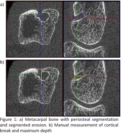
Methods: In the MCP joints of the second to forth digit of 18 patients 80 slices distally and 242 slices proximally to the surface of the third metacarpal head were scanned (XtremeCT, Scanco Switzerland; isotropic voxel size 82µm). Erosions were quantified as follows: After segmenting the periosteal surface the user identified each erosion by manually placing a seed point. The erosions were then automatically segmented by a 3D level-set algorithm (Fig. 1a) with the option of operator corrections. Erosion volume (Vol), surface area (SA), and sphericity (SP), a parameter describing the shape deviation from a perfect sphere, were determined. In addition manual measurements were carried out. Here the erosion volume was approximated by a half-ellipsoid constructed from the surface area of the cortical break and the maximum erosion depth perpendicular to it (Fig. 1b). In order to compare both methods the lengths measurements obtained from the manual technique were also determined during the automated 3D analysis.
Results: 32 erosions were assessed in the 18 datasets with a mean/min/max Vol of 9.66mm³/0.37mm³/54.7mm³ (automated 3D analysis). Inter-operator precision errors (3 operators, root mean squares coefficients of variation (RMSCV)/RMS standard deviation (RMSSD)) were 7.8%/0.8 mm³, 10.4%/3.8 mm2 and 4.9%/0.02 for Vol, SA and SP, respectively. Excluding 18 erosions, in which operator interactions were performed, decreased the errors by about a factor of 3. Correlation between the manual analysis and the length measurements obtained from the automated 3D analysis was r = 0.87, however, correlation with the Vol obtained from the full 3D analysis was r = 0.39, indicating that a simplistic approximation of erosion volume may not capture the full shape information.
Conclusion: We developed a new precise full 3D characterization of bone erosions that may help improving the assessment of disease activity and treatment efficacy. Precision errors depend on the degree of user interaction that may be necessary to correct the automated segmentation, which is more frequent in erosions with large cortical breaks. Manual measurements are less impacted by image quality, such as motion artifacts; however, the approximation of erosion volume by a half-ellipsoid underestimates the true erosion volume. The clinical evaluation of this method is currently being performed.
Disclosure:
D. Toepfer,
None;
S. Finzel,
None;
O. Museyko,
None;
K. Engelke,
None;
G. A. Schett,
None.
« Back to 2012 ACR/ARHP Annual Meeting
ACR Meeting Abstracts - https://acrabstracts.org/abstract/segmentation-and-quantification-of-bone-erosions-in-the-hands-of-patients-with-rheumatoid-arthritis-using-high-resolution-computed-tomography/

