Session Information
Session Type: Abstract Submissions (ACR)
Background/Purpose: Radiographic joint space width (JSW) assessment, a surrogate for cartilage assessment, is the standard for structure modification trials of osteoarthritis (OA). However the subchondral bone is integral to OA progression and modern image analysis techniques allow accurate, automated identification of bone in MR images. The objective of this study was a comparison of the sensitivity of 3D bone area measures in MR images with minimum medial JSW in radiographs in all subjects in the Osteoarthritis Initiative dataset with definite medial OA over a 2 year period, representative of a typical OA clinical trial
Methods: 828 subjects with medial OA and MR images at baseline, 12 and 24 months, and available radiograph scoring were selected from the Osteoarthritis Initiative dataset; medial OA was defined as KL≥2 and presence of medial osteophytes. Femur, tibia and patella bones were automatically segmented from MRIs using active appearance models1. Anatomical areas were automatically identified within the model2 and were measured at each time-point. All regions of the articulating surface of the femur, tibia and patella were included in the analysis. Minimum medial joint space width (minMJSW) was provided by the OAI, using a semi-automated software method3. Sensitivity of both methods to change was assessed using the standardised response mean (mean/SD of change)
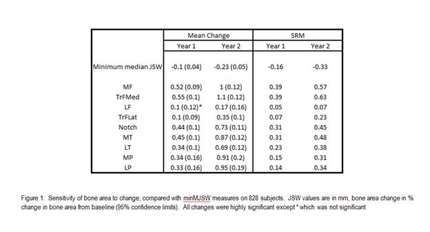
Results: Mean age (SD) of the case group was 62 years (8.8); BMI 29.7(4.8); KL 2.55(0.65); 35% females. MinMJSW showed significant change at 12 and 24 months with SRM values of -0.16 and -0.33. Change in bone area was significant in all regions for all time-points, except the lateral femur at 12 months (see Figure 1). Medial femur compartments provided the greatest sensitivity to change, with SRM values typically twice those of minMJSW. Tibial compartments and the notch of the femur also showed higher SRM figures than minMJSW, while patellar compartments had comparable SRMs to the radiographic measure. Only the lateral femoral compartments had lower SRMs than the minMJSW method.
Conclusion: Change in bone area was more sensitive than the minMJSW method in a number of knee compartments, particularly in the medial femur and tibia. Measurement of bone change provided a more responsive tool for monitoring OA progression in a cohort selected for typical OA trial characteristics.
References:
1 Cootes, T. F, G. J. Edwards, and C. J. TaylorIEEE Trans Patt Anal Mach Intell 23.6 (2001): 681-85.
2 Williams, T. G., et al. Br.J.Radiol. 83.995 (2010): 940-48.
3 Duryea J, Li J, Peterfy CG, Gordon C, Genant HK. Med Phys. 2000 Mar: 27(2): 580-91
Disclosure:
M. A. Bowes,
Imorphics Ltd,
1,
Imorphics Ltd,
3;
C. B. Wolstenholme,
Imorphics Ltd,
3,
Imorphics Ltd,
1;
D. Hopkinson,
None;
G. R. Vincent,
Imorphics Ltd,
3,
Imorphics Ltd,
1;
P. G. Conaghan,
None.
« Back to 2012 ACR/ARHP Annual Meeting
ACR Meeting Abstracts - https://acrabstracts.org/abstract/changing-osteoarthritis-treatment-assessment-paradigms-subchondral-bone-is-a-more-responsive-measure-of-progression-than-the-current-radiographic-standard/

