Session Information
Session Type: Abstract Submissions (ACR)
Background/Purpose: The imaging arm of the ASAS axial spondyloarthritis (SpA) criteria requires the presence of sacroiliitis on MRI or radiographs. In patients (pts) with non-radiographic axial SpA (nr-axSpA), there may be inflammation along the spine in the absence of sacroiliac joint (SIJ) inflammation on MRI. This analysis evaluated the existence of spinal inflammation on MRI at baseline (BL) in nr-axSpA pts with and without inflammation in the SIJs on MRI.
Methods: ABILITY-1 is an ongoing multicenter, randomized, controlled trial of adalimumab vs. placebo in pts with nr-axSpA classified using the ASAS axial SpA criteria, who had an inadequate response, intolerance to, or contraindication for NSAIDs. MRI of the SIJ and spine performed at BL were centrally scored using the SPARCC method (6-DVU method for the spine) by 2 independent readers blinded to the treatment codes. Mean scores of the readers were used. SPARCC score ≥2 for either the SIJ or spine was used as the operational definition of positive MRI evidence of inflammation. For these analyses, all pts were combined, independent of randomization.
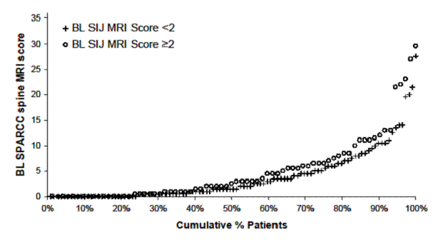
Results: Mean symptom duration of the study population (N=185) was 10 yrs. At BL, 48% of pts were reported by the local investigator to have past or present MRI evidence of sacroiliitis as required by the ASAS axial SpA criteria. Of pts with available BL SPARCC scores, 40% had a BL SIJ score ≥2 and 52% had a BL spine score ≥2. Of the pts with BL SPARCC SIJ score <2, 49% had evidence of spinal inflammation (BL SPARCC spine score ≥2). Comparison of BL disease characteristics based on BL spine and SIJ scores <2 vs. ≥2 were generally comparable except for a greater proportion of males among those with spine and SIJ scores ≥2, and younger age and shorter symptom duration among those with spine and SIJ scores <2. The cumulative probability plot (figure) shows a similar distribution of SPARCC spine scores regardless of presence or absence of SIJ inflammation on MRI. The most frequently involved DVUs with bone marrow edema were in the lower thoracic and lumbar spine.
Conclusion: Assessment by experienced readers shows that spinal inflammation on MRI may be observed in half of nr-axSpA pts without SIJ inflammation on MRI. MRI of both sites might be of value when evaluating pts with nr-axSpA. These data in pts with long-standing disease need to be confirmed in pts with shorter disease duration.
Figure
Disclosure:
D. van der Heijde,
Abbott Laboratories; Amgen; AstraZeneca; BMS; Centocor: Chugai; Eli-Lilly; GSK; Merck; Novartis; Pfizer; Roche; Sanofi-Aventis; Schering-Plough; UCB; Wyeth,
5,
Abbott Laboratories; Amgen; AstraZeneca; BMS; Centocor: Chugai; Eli-Lilly; GSK; Merck; Novartis; Pfizer; Roche; Sanofi-Aventis; Schering-Plough; UCB; Wyeth,
2,
Imaging Rheumatology,
4;
J. Sieper,
Abbott, Merck, Pfizer, and UCB,
2,
Abbott, Merck, Pfizer, and UCB,
5,
Abbott, Merck, Pfizer, and UCB,
8;
W. P. Maksymowych,
Abbott, Amgen, BMS, Eli-Lilly, Janssen, Merck, and Pfizer,
2,
Abbott, Amgen, BMS, Eli-Lilly, Janssen, Merck, and Pfizer,
5;
M. A. Brown,
Abbott Laboratories,
5;
S. S. Rathmann,
Abbott Laboratories,
3,
Abbott Laboratories,
1;
A. L. Pangan,
Abbott Laboratories,
3,
Abbott Laboratories,
1.
« Back to 2012 ACR/ARHP Annual Meeting
ACR Meeting Abstracts - https://acrabstracts.org/abstract/spinal-inflammation-in-the-absence-of-si-joint-inflammation-on-mri-in-patients-with-active-non-radiographic-axial-spondyloarthritis/

