Session Information
Date: Sunday, November 13, 2016
Title: Osteoporosis and Metabolic Bone Disease – Clinical Aspects and Pathogenesis - Poster
Session Type: ACR Poster Session A
Session Time: 9:00AM-11:00AM
Background/Purpose:
To study and summarize the clinical features of tumor-induced osteomalacia(TIO) misdiagnosed as spondyloarthritis(SpA), aiming to explore the reasons of misdiagnosis and raise the correctness of TIO diagnosis.
Methods: A total of 18 TIO patients misdiagnosed as SpA from March 2002 to January 2016 in Department of Rheumatology of Chinese PLA General Hospital were enrolled. The clinical manifestations, laboratory examinations data, imaging features, Localization of tumors and histological characteristics were analyzed.
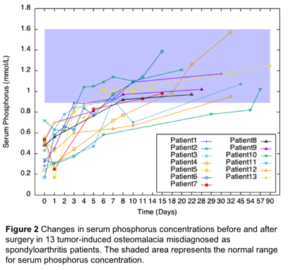
Results: 18 patients were included (11 males and 7 females) with a median age of 33 years (range 19–60). The mean disease duration was 3.8 years (range 0.17 to 20 years). All patients presented with persistent low back pain. The pain could not be relieved by rest or exercise, all patients had a history of taking nonsteroidal anti-inflammatory drugs and showed no obviously improvement. The inflammatory markers such as erythrocyte sedimentation rate and C-reactive protein were usually normal. The level of serum calcium was normal or slightly lower, nevertheless, all patients had hypophosphatemia and increased level of alkaline phosphatase. Sacroiliac joint lesions were found in X-ray, CT or MRI, however the lesions in sacrum or ilium were predominant rather than in joints. Abnormal bone imaging in ribs, long bones and soft tissues in addition to joints could be detected by bone scintigraphy. And the technetium-99m octreotide scintigraphy (Figure 1a) was completed to detect the causative neoplasms, then targeted images with radiographs (Figure 1b), CT or MRI were used for further localization of the tumors. Successful localization of tumors was followed by surgical resection (13 of these 18 patients) and analysis by pathology studies. The pathology results indicated that the majority (46.2 %,6 of 13) were characterized as phosphaturic mesenchymal tumors (PMTs). After tumor resection, serum phosphorus levels normalized in all 13 surgical patients after a mean of 16 days (range 3-57 days), and Clinical symptoms were alleviated within 3 months (Figure 2).
Conclusion: TIO can be easily misdiagnosed as SpA because of the atypical clinical features and imaging changes of sacroiliac joint, however, the laboratory findings and response to the NSAIDs of the two conditions are different. Considering the complete surgical resection leading to normalization of parameters in laboratory tests and relief of symptoms of TIO patients, prompt diagnosis and surgical treatment is extremely essential. Comprehensive laboratory examinations and the technetium-99m octreotide scintigraphy can contribute to the timely diagnosis of TIO.
To cite this abstract in AMA style:
Sui N, Zhu J, Sun F. Clinical Analysis of Tumor-Induced Osteomalacia Misdiagnosed As Spondyloarthritis: A Report of 18 Cases [abstract]. Arthritis Rheumatol. 2016; 68 (suppl 10). https://acrabstracts.org/abstract/clinical-analysis-of-tumor-induced-osteomalacia-misdiagnosed-as-spondyloarthritis-a-report-of-18-cases/. Accessed .« Back to 2016 ACR/ARHP Annual Meeting
ACR Meeting Abstracts - https://acrabstracts.org/abstract/clinical-analysis-of-tumor-induced-osteomalacia-misdiagnosed-as-spondyloarthritis-a-report-of-18-cases/


