Session Information
Date: Monday, November 9, 2015
Title: Systemic Lupus Erythematosus - Clinical Aspects and Treatment Poster Session II
Session Type: ACR Poster Session B
Session Time: 9:00AM-11:00AM
Background/Purpose: Cardiovascular disease (CVD) is a
leading cause of death in systemic lupus erythematosus (SLE) and in rheumatoid
arthritis (RA). Longer corrected QT segments (QTc) and a high prevalence of
repolarization abnormalities have been recently described in the electrocardiograms
(EKG) of SLE patients. In addition, both QTc prolongation and ST-T changes have
been associated with increased cardiovascular mortality in the general
population. Our objective was to evaluate QTc duration and ST-T changes in a
tertiary lupus center and compare it with a cohort of RA patients without CVD.
Methods: Fifty SLE patients meeting ACR
classification criteria were cross-sectionally evaluated and compared with 139
RA patients without known CVD. Demographics, disease characteristics, disease
activity, and comorbidities were ascertained. Linear and logistic regression models
were used to assess the associations with QTc duration and ST-T changes,
adjusting for CVD risk factors.
Results:
SLE patients (38
±14 years old, 92% females, 73% Hispanic) had a median disease duration of 8
years and median SLEDAI-2K of 6. Forty-four percent had a history of lupus nephritis,
62% were on antimalarials, and 52% and 30% were SSA and SSB antibody positive,
respectively. RA patients (59 ± 8 years old, 61% females, 87% white) had a
median disease duration of 8 years, a mean DAS28-CRP of 3.6, 68% were anti-CCP
antibody positive, and 48% were on biologics. SLE patients were more likely to
be smokers (24% vs. 9%), and 7 (14%) had known CVD. There was no difference in
the prevalence of hypertension, diabetes, aspirin or statin use between the two
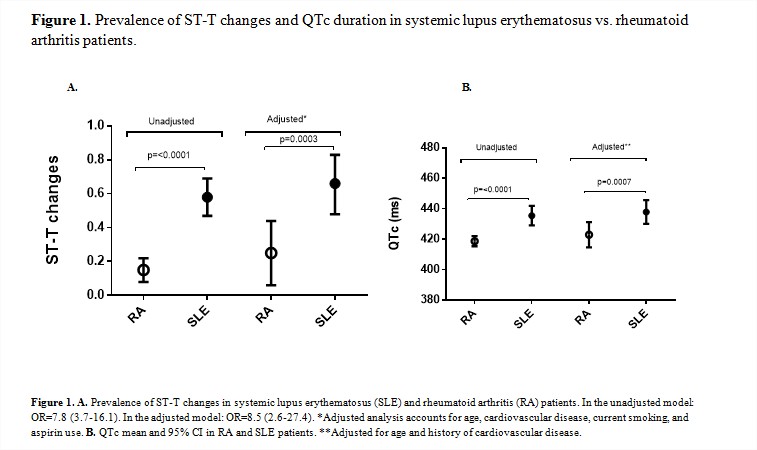
groups (Table 1). The prevalence of ST-T changes was higher in SLE vs. RA (58%
vs. 15%, respectively; p<0.0001) representing an 8-fold higher odds. After
adjusting for age, smoking, CVD, and aspirin use, the difference remained
statistically significant (OR=8.5; p=0.0003). The QTc duration was
significantly longer in SLE vs. RA patients in the univariable and in the
adjusted analyses (435 vs. 418 ms, p=<0.0001; and 438 vs. 423 ms, p=0.0007,
respectively) (Figure 1). Similar results were seen when limiting the analysis
to only those without CVD. In SLE patients no specific disease characteristics
were associated with these EKG changes. In the RA group, biologics were
protective for the occurrence of ST-T changes.
Conclusion: More frequent ST-T changes and a
longer QTc duration were seen in SLE when compared to RA patients. Longitudinal
studies to evaluate the progression of these EKG changes and their risk for
life-threatening arrhythmias and cardiovascular death in these patients are
needed.
To cite this abstract in AMA style:
Geraldino-Pardilla L, Gartshteyn Y, Giles JT, Perez T, Askanase AD, Bathon JM. Comparison of Electrocardiographic ST-T Changes and QTc Duration in Patients with Systemic Lupus Erythematosus and Rheumatoid Arthritis [abstract]. Arthritis Rheumatol. 2015; 67 (suppl 10). https://acrabstracts.org/abstract/comparison-of-electrocardiographic-st-t-changes-and-qtc-duration-in-patients-with-systemic-lupus-erythematosus-and-rheumatoid-arthritis/. Accessed .« Back to 2015 ACR/ARHP Annual Meeting
ACR Meeting Abstracts - https://acrabstracts.org/abstract/comparison-of-electrocardiographic-st-t-changes-and-qtc-duration-in-patients-with-systemic-lupus-erythematosus-and-rheumatoid-arthritis/


