Session Information
Date: Monday, November 9, 2015
Title: Spondylarthropathies and Psoriatic Arthritis - Comorbidities and Treatment Poster II
Session Type: ACR Poster Session B
Session Time: 9:00AM-11:00AM
Background/Purpose:
There is a well-documented association between inflammatory bowel disease (IBD)
and ankylosing spondylitis (AS). The hallmark feature of AS is the presence of
sacroiliitis that has traditionally been diagnosed using an AP view of the
pelvis on X-ray. Many IBD patients receive supine and erect abdominal X-rays in
the management of their bowel disease and it is not known whether these films
can be used in the diagnosis of AS.
Many experts
agree that CT scan is a superior imaging modality for the diagnosis of
sacroiliitis though until recently there was no definition of what constituted
a positive CT. Recently we have described a standardized scoring system for the
diagnosis of sacroiliitis which is anchored in the modified New York criteria
(mNY). To date, there has not been a study assessing the diagnostic utility of
abdominal X-rays for the diagnosis of spondyloarthritis in IBD patients.
We aim to
compare the reproducibility and test characteristics of abdominal X-rays in
detecting CT-positive sacroiliitis in a IBD cohort.
Methods: In a
previous study, we identified IBD patients who had a CT scan of their
abdomen/pelvis from one IBD clinic. CT scans were read by 2 blinded readers and
49 patients were identified as having sacroiliitis according to a recently
validated scoring system. Of these 49 patients, 33 were found to have abdominal
X-rays done within 1 year of their CT scan.
The 33
patients included in the study were matched by age, gender, and IBD subtype to
control IBD patients (n=33). X-rays were randomized and read by two experienced
blinded readers according to modified New York grade. Discordant results were
settled by a third reader. Patients were classified as having diagnostic or
non-diagnostic SI joints and inter-reader reliability as well as the prevalence
of specific features of sacroiliitis was determined.
Results: The
median age for IBD patients was 35 years (range 17-75) and 64% were male.
Within the CT positive group, 21/33 (64%) were scored as having diagnostic
X-rays according to mNY grading system. Amongst the CT-negative control group, X-rays
identified sacroiliitis in 3/33 (9%) patients. Inter-reader reliability for diagnosis
of sacroiliitis using X-rays was good (κ=0.659)
which was similar to that found with CT scan (κ=0.697). Cramér’s V correlation between the two
imaging modalities revealed an r=0.647. Chi squared test was used to compare
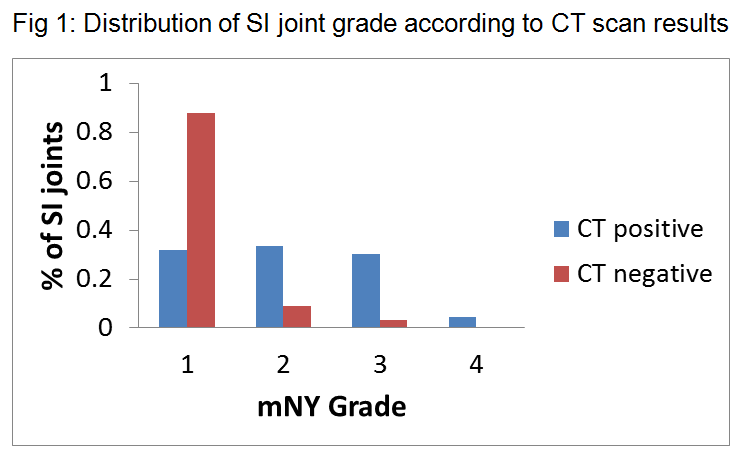
the two nominal variables and showed a p value <0.0001. Fig 1 shows the distribution
of SI joint grade according to CT scan result.
Conclusion: Abdominal
X-ray and CT scan have a similar inter-reader reproducibility for the diagnosis
of sacroiliitis. Abdominal X-ray is able to detect the majority of patients
with CT-positive sacroiliitis; however, 36% of CT-positive patients would not
have been identified. These results need to be further correlated with clinical
context to confirm a diagnosis of spondyloarthritis.
To cite this abstract in AMA style:
Chan J, Sari I, Omar A, Silverberg MS, Salonen D, Inman RD, Haroon N. Abdominal X-Rays Can Reliably Detect the Majority of CT Positive Sacroiliitis in an IBD Cohort [abstract]. Arthritis Rheumatol. 2015; 67 (suppl 10). https://acrabstracts.org/abstract/abdominal-x-rays-can-reliably-detect-the-majority-of-ct-positive-sacroiliitis-in-an-ibd-cohort/. Accessed .« Back to 2015 ACR/ARHP Annual Meeting
ACR Meeting Abstracts - https://acrabstracts.org/abstract/abdominal-x-rays-can-reliably-detect-the-majority-of-ct-positive-sacroiliitis-in-an-ibd-cohort/

